. 2006 Sep;4(9):e276.
doi: 10.1371/journal.pbio.0040276.

Disruption-induced mucus secretion: repair and protection

Affiliations

Disruption-induced mucus secretion: repair and protection

K Miyake et al. PLoS Biol. 2006 Sep.

Abstract

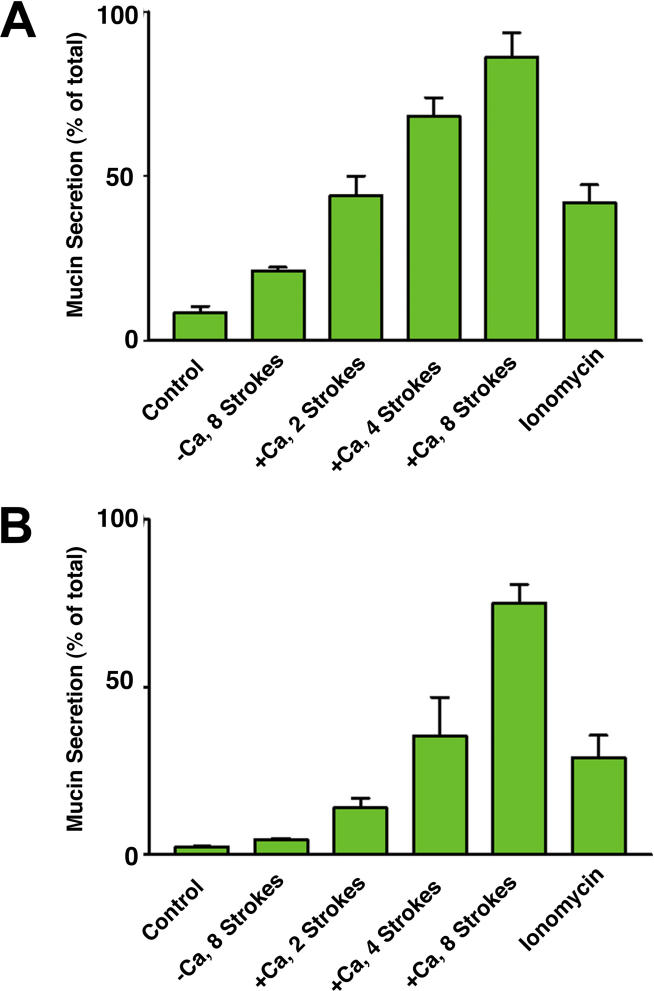
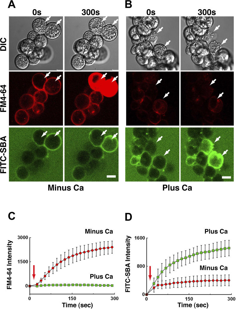
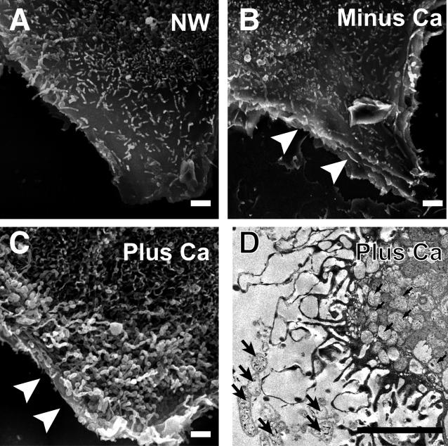
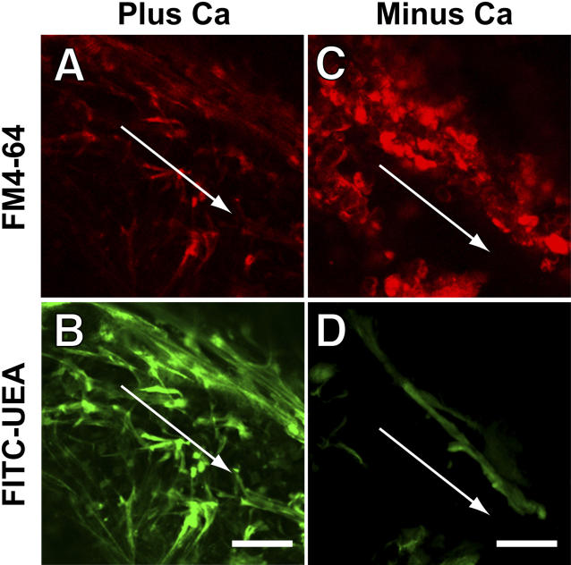
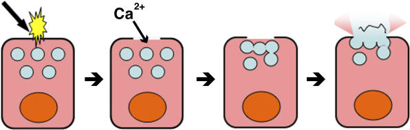
When a cell suffers a plasma membrane disruption, extracellular Ca(2+) rapidly diffuses into its cytosol, triggering there local homotypic and exocytotic membrane fusion events. One role of this emergency exocytotic response is to promote cell survival: the internal membrane thus added to the plasma membrane acts as a reparative "patch." Another, unexplored consequence of disruption-induced exocytosis is secretion. Many of the cells lining the gastrointestinal tract secrete mucus via a compound exocytotic mechanism, and these and other epithelial cell types lining the digestive tract are normally subject to plasma membrane disruption injury in vivo. Here we show that plasma membrane disruption triggers a potent mucus secretory response from stomach mucous cells wounded in vitro by shear stress or by laser irradiation. This disruption-induced secretory response is Ca(2+) dependent, and coupled to cell resealing: disruption in the absence of Ca(2+) does not trigger mucus release, but results instead in cell death due to failure to reseal. Ca(2+)-dependent, disruption-induced mucus secretion and resealing were also demonstrable in segments of intact rat large intestine. We propose that, in addition to promoting cell survival of membrane disruptions, disruption-induced exocytosis serves also the important protective function of liberating lubricating mucus at sites of mechanical wear and tear. This mode of mechanotransduction can, we propose, explain how lubrication in the gastrointestinal tract is rapidly and precisely adjusted to widely fluctuating, diet-dependent levels of mechanical stress.


Conflict of interest statement

Competing interests. The authors have declared that no competing interests exist.

Figures

Figure 1
Figure 1. Three Lectins Strongly Stain the Intracellular Content of Gastric Surface Mucous Cells
Frozen sections of the fundic portion of the mouse stomach were stained with rhodamine-labeled SBA (A), WGA (B), or UEA1 (C). Note staining of the surface mucous (SM) cells, which are the mucus-producing population of the gastric epithelium. Primary cultures of gastric surface mucous cells (asterisks) were also stained with SBA (D), WGA (E), or UEA1 (F). Confocal imaging reveals labeling of a spherical, large (~ 1-μm diameter), abundant organelle, the mucus granule. Bars represent 10 μm.
Figure 2
Figure 2. Shear Stress Induces Ca2+-Dependent Secretion of Mucus by Cultured Gastric Mucous Cells
Cells were taken up into and ejected from (one “stroke”) a syringe operating under constant pressure in the presence of 1.5 mM Ca2+ (+Ca), or were not so treated (Control). One group of cells was wounded by syringing in the absence of extracellular Ca2+ (−Ca), and another was incubated with a Ca2+ ionophore (Ionomycin, 1 μM, 10 min). The medium conditioned by these cells was then harvested and the content of mucus present quantitated in an ELLA assay employing either WGA (A) or UEA (B) as the mucus ligand. All treatments, except wounding in the absence of Ca2+ (−Ca, eight strokes), gave mean (± standard error of the mean) values that differed significantly from the control, p ≤ 0.001 (A), p ≤ 0.01 (B), Kruskal-Wallis test.
Figure 3
Figure 3. Cell Resealing and Mucus Secretion Are Ca2+-Dependent, Concurrent Events in Surface Mucous Cells
(A) A cluster of gastric mucous cells was imaged by confocally before (0 s) and after (300 s) wounding of two cells (arrows) with a laser in the absence of Ca2+ (Minus Ca). Shown are differential interference contrast (DIC), and fluorescence images of FM4–64 dye (red channel), which stains the cytoplasmic membranes of cells that fail to reseal, and of FITC-SBA (green channel), which stains extracellular mucus. Note that strong cytoplasmic staining with FM4–64 is seen 300 s after wounding, whereas no detectable increase in surface staining with FITC-SBA is observed at this time point. (B) A cluster of gastric mucous cells wounded and imaged as in (A) but in the presence of 1.5 mM Ca2+ (Plus Ca). Note that very little cytoplasmic staining with FM4–64 is seen 300 s after wounding, whereas strong surface staining with FITC-SBA is observed at this time point. (C) Entry of FM 4–64 dye into cell cytoplasm was monitored over time after laser wounding (arrow). In the presence of extracellular Ca2+ (Plus Ca), a small amount of dye entry is detectable only during the first 20–30 s post-wounding, indicating that resealing was completed within this time frame. In the absence of extracellular Ca2+ (Minus Ca), entry of dye continues until, at approximately 100 s, internal membranes reach a saturation point. Resealing failed in these cells (n = 3). Bars indicate the standard error of the mean. (D) Surface staining intensity of the cells with FITC-SBA monitored over time after laser wounding (red arrow). In the presence of extracellular Ca2+ (Plus Ca2+), an increase in surface staining is detectable at 50 s post-wounding and continues throughout the time course of the experiment, indicating that, concurrently with resealing, mucus was being exocytosed. In the absence of Ca2+ (Minus Ca2+), by contrast, only a slight increase in staining was observed (n = 3). Bars indicate the standard error of the mean.
Figure 4
Figure 4. Cells That Survive a Plasma Membrane Disruption Are Depleted of Intracellular Mucus
(A) MKN28 cell monolayer substrata were scratched with the tip of a needle, denuding cells and wounding many of those bordering on the denudation site. Those cells that resealed the plasma membrane disruptions that were thus created trap in their cytoplasm the Texas red-labeled (TRDx) dextran present during the scratch injury, and their cytosol is consequently fluorescent (arrows). (B) Images of the same cells after staining of intracellular mucus with FITC-WGA. Note that those cells heavily labeled with the TRDx-dextran, which suffered and repaired large plasma membrane disruptions, apparently stain more lightly with the FITC-WGA. (C) Flow cytofluorometic analysis of FITC-WGA staining in MKN28 populations that were wounded by syringing (W) or were undisturbed (NW) prior to intracellular staining of mucus with FITC-WGA. Note the downward shift in population fluorescence of the wounded relative to the undisturbed population. (D) The mean value of wounded (W) and undisturbed (NW) population fluorescence as measured by flow cytofluorometry (n = 3; p < 0.05). Bars represent 20 μm.
Figure 5
Figure 5. Mucus-Producing Cells Exhibit a Ca2+-Dependent Change in Surface Architecture following Wounding
Subconfluent monolayers of cultured MKN28 cells were wounded by scratching the glass substratum with a needle tip, and then prepared for scanning electron microscopy. (A) Scanning electron micrograph of an undisturbed cell (NW) not bordering on a scratch site. (B) Scanning electron micrograph of a cell bordering on a scratch site made in the absence of extracellular Ca2+ (Minus Ca). The arrowheads mark a site of plasma membrane discontinuity, presumably a disruption that failed to reseal. (C) Scanning electron micrograph of a cell bordering a wound site made in physiological Ca2+ (Plus Ca). Arrowheads mark the presumptive plasma membrane disruption site. Note the dramatic difference (from cells in [A] and [C]) in surface architecture adjacent to this presumptive membrane disruption site, which includes numerous villus-like projections of membrane, and spherical profiles, presumably the exocytosed content of mucus granules. (D) Transmission electron micrograph of a cell bordering on a wound site made in physiological Ca2+. Entry of horseradish peroxidase, present extracellularly while making the scratch, results locally in the prominent dark labeling of villi and other sub-plasma membrane spaces, and marks this location as one at or nearby the disruption site. Note the numerous, microvillar extensions and the presence of mucus granules both within the cell (arrows) and decorating its surface. Bars represent 2 μm.
Figure 6
Figure 6. Ca2+-Dependent Secretion of Mucus, and Resealing, Can Be Observed in an Excised Segment of Colon
A segment of excised colon was scratched (the arrows mark the approximate sites) in the presence (Plus Ca) and absence (Minus Ca) of physiological Ca2+. FM4–46 dye, a marker for cells that failed to reseal, and FITC-UEA, a marker of surface mucus, were added 3 min later. Confocal images of the segment were then immediately acquired of the FM4–64 (A) and (C) and FITC-UEA (B) and (D) staining. As indicated by relative FM4–64 staining intensities, an increased incidence of resealing failure occurred in the absence of Ca2+. As indicated by the relative FITC-UEA staining intensities, on the other hand, a dramatically decreased incidence of mucus secretory events occurred in the absence of Ca2+. Bars represent 50 μm.
Figure 7
Figure 7. Coupling of Repair and Protective Mucus Secretion
Mechanical or other stressors produce a plasma membrane disruption in a mucous cell lining the GI tract. Ca2+ enters through the disruption. Homotypic fusion of granules with one another is triggered by this signal. Exocytotic fusion of the homotypic fusion product with the plasma membrane surrounding the defect site completes repair and liberates mucus into the extracellular environment. Some cytoplasmic components and membrane are also liberated (see text for explanation).

Comment in

  • The path to digestion is paved with repair.
    Underwood J. Underwood J. PLoS Biol. 2006 Sep;4(9):e307. doi: 10.1371/journal.pbio.0040307. Epub 2006 Aug 22. PLoS Biol. 2006. PMID: 20076638 Free PMC article. No abstract available.

References

    1. McNeil PL, Steinhardt RA. Plasma membrane disruption: Repair, prevention, adaptation. Annu Rev Cell Dev Biol. 2003;19:697–731. - PubMed
    1. McNeil PL, S, Ito S. Gastrointestinal cell plasma membrane wounding and resealing in vivo. Gastroenterology. 1989;96:1238–1248. - PubMed
    1. Forstner G. Signal transduction, packaging and secretion of mucins. Annu Rev Physiol. 1995;57:585–605. - PubMed
    1. Bickel M, Kauffman GL., Jr Gastric gel mucus thickness: Effect of distention, 16,16-dimethyl prostaglandin e2, and carbenoxolone. Gastroenterology. 1981;80:770–775. - PubMed
    1. Enss ML, Schmidt-Wittig U, Honer K, Kownatzki R, Gartner K. Mechanical challenge causes alterations of rat colonic mucosa and released mucins. Alterations of mucosa and mucins. J Exp Anim Sci. 1994;36:128–140. - PubMed

Publication types