. 2006 Aug 25;2(8):e134.
doi: 10.1371/journal.pgen.0020134. Epub 2006 Jul 13.

Overlapping functions of argonaute proteins in patterning and morphogenesis of Drosophila embryos

Affiliations

Overlapping functions of argonaute proteins in patterning and morphogenesis of Drosophila embryos

Wibke J Meyer et al. PLoS Genet. .

Abstract

Argonaute proteins are essential components of the molecular machinery that drives RNA silencing. In Drosophila, different members of the Argonaute family of proteins have been assigned to distinct RNA silencing pathways. While Ago1 is required for microRNA function, Ago2 is a crucial component of the RNA-induced silencing complex in siRNA-triggered RNA interference. Drosophila Ago2 contains an unusual amino-terminus with two types of imperfect glutamine-rich repeats (GRRs) of unknown function. Here we show that the GRRs of Ago2 are essential for the normal function of the protein. Alleles with reduced numbers of GRRs cause specific disruptions in two morphogenetic processes associated with the midblastula transition: membrane growth and microtubule-based organelle transport. These defects do not appear to result from disruption of siRNA-dependent processes but rather suggest an interference of the mutant Ago2 proteins in an Ago1-dependent pathway. Using loss-of-function alleles, we further demonstrate that Ago1 and Ago2 act in a partially redundant manner to control the expression of the segment-polarity gene wingless in the early embryo. Our findings argue against a strict separation of Ago1 and Ago2 functions and suggest that these proteins act in concert to control key steps of the midblastula transition and of segmental patterning.


Conflict of interest statement

Competing interests. The authors have declared that no competing interests exist.

Figures
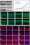
Figure 1
Figure 1. dop1 Affects Cell Formation in the Early Embryo
(A–C) Membrane extension is strongly reduced in dop1 embryos. (A, A′) Frames taken from a video-sequence of a wild-type embryo. In the wild-type, membranes grow slowly for the first 30 min of cycle 14 interphase [slow phase (A)], and then growth speeds up considerably [fast phase (A′)]. (B, B′) dop1 mutant embryo from a video-sequence at corresponding time points. The shape of the nuclei (circles) and the extent of furrow progression are indicated (bars). (C) The progression of the cleavage furrow was plotted against the time after the beginning of mitotic cycle 14. Note that compared to the wild-type furrow progression is significantly slower in embryos derived from homozygous (dop1/dop1) or hemizygous [dop1/Df(3L)XG9] dop1 females, within the first 30 min of cellularization. In dop1 homozygotes, membranes advanced 0.07 μm/min during slow phase, compared to 0.25 μm/min in the wild-type. In hemizygous dop1 embryos, membrane formation was even more severely impaired; it was slower during both slow and fast phase. (D, E) Immunostaining of the endogenous membrane protein Neurotactin (Nrt; green) and DNA (blue) in wild-type (D) and dop1 (E) mutant embryos during progression of cell formation (from left to right panel). While Nrt associates with the egg cortex in both cases, Nrt positive cleavage membranes in dop1 mutant embryos are absent until fast phase. (F–I) Immunostaining with Slam antibodies (red) indicates the presence of furrow canals in wild-type (F, H) and dop1 (G, H) mutant embryos (F, G: optical cross section; H, I: tangential optical section) at progressively older stages of cell formation. (F, H) In the wild-type, formation of the furrow canal can be seen as a regular array of loop-like structures beginning with nuclear elongation. (G, I) In dop1 mutant embryos, Slam localization does not resolve into this regular array and forms an unevenly distributed network apical to the nuclei. (J–M) Immunostaining with Arm antibodies (red) indicates the positioning of the basal junctions; two timepoints are shown in each panel: beginning of slow phase to the left and beginning of fast phase to the right. (J, L) In the wild-type, Arm accumulates adjacent to the furrow canals and forms a honeycomb pattern as seen in surface view. (K, M) In dop1 mutant embryos, Arm remains apical and does not accumulate in basal junctions. Fixed embryos were staged by the extent of nuclear elongation, as described by Lecuit et al. [30].
Figure 2
Figure 2. dop1 Compromises Polarized Microtubule-Based Transport
(A, B) During germband extension, the periphery of dop1 embryos (B) is much more transparent than that of wild-type embryos (A). (C, D) Lipid droplets (green) were stained with the droplet-specific fluorescent dye Nile Red, and their distribution was recorded by epifluorescence microscopy. In the wild-type, lipid droplets are found throughout the periphery. In the mutant, lipid droplets are accumulated around the central yolk. (E–H) Males homozygously deleted for halo were crossed to dop1 heterozygous (E, G) or homozygous (F, H) females to generate embryos with reduced halo expression. In these embryos, droplet distribution was assessed by overall transparency (E, F) or staining for the regulator Klar (green) (G, H). In late cycle 14, embryos from dop1 homozygous mothers have a more transparent periphery and tighter basal accumulation of Klar puncta. (I, K) Overall expression and distribution of Klar (green) is very similar in wild-type (I) and ago2dop1 embryos (K). (L) In centrifuged early embryos, lipid droplets accumulate in a distinct layer, just above nuclei (blue). In centrifuged dop1 embryos, Klar (green) is highly enriched in the droplet layer, just as in the wild-type [36], indicating that it is physically associated with the droplets. Scale bars represent 200 μm in (A) and 80 μm in (L). (G–L) Nuclei (blue) were labeled with Hoechst 33258.
Figure 3
Figure 3. Genetic and Molecular Analysis of the ago2dop Mutations
(A) Genetic characterization of the dop genomic region. Deletion mapping identified the cytological interval 71D1-E1 as the region uncovering the dop locus. Black bars represent the deleted regions. Df(3L)BK10 represents the deletion that was used in the initial screen for female-sterile mutations by Galewsky and Schulz [25]. The distal breakpoint of Df(3L)XG9 is to the right of the mex1 gene (based on PCR mapping) and the proximal breakpoint is to the left of CG7739 (based on complementation of l(3)s1754 and PCR probing for CG7739). (B) Genomic organization of the 71D1 to 71E3 region as predicted from the release 3 of the annotated genomic sequence by the Berkeley Drosophila Genome Project. The 45-kb region contains six predicted genes and three P-element-insertions. The P-insertions P(l(3)s1754), P(l(3)03576, and P{EP}EP3417 were utilized to produce small deletions by male recombination (unpublished data).
Figure 4
Figure 4. Requirement of ago2 and ago1 for siRNA-Mediated RNAi
Effects of mutations on siRNA-mediated RNAi were assayed using a UAS construct that reduces the function of the cell death inhibitor DIAP1 by expression of dsRNA (DIAP1RNAi: GMR::Gal4,UAS::DIAP1RNAi) [38]. DIAP1RNAi in the compound eye leads to the loss of cells and reduces the normal size of the eye. Scanning EM micrographs of compound eyes of 2- to 4-d-old females of the following genotypes are indicated: (A) wild-type compound eye; (B) GMR::Gal4, UAS::DIAP1RNAi/CyO; (C) GMR::Gal4, UAS::DIAP1RNAi/dcr-2L811fsX; (D) GMR::Gal4, UAS::DIAP1RNAi/CyO; ago251B/ago251B; (E) GMR::Gal4, UAS::DIAP1RNAi/CyO; ago2dop1/TM6; (F) GMR::Gal4, UAS::DIAP1RNAi/CyO; ago2dop1/ago2dop1; (G) GMR::Gal4, UAS::DIAP1RNAi; ago2dop1/Df(3L)XG9; (H) GMR::Gal4, UAS::DIAP1RNAi/ago1K08121; (I) GMR::Gal4, UAS::DIAP1RNA/ago1K08121; ago2dop1/TM6; and (J) GMR::Gal4, UAS::DIAP1RNAi/ago1K08121; ago2dop1/ago2dop1.
Figure 5
Figure 5. Genetic Interaction of ago2dop1 with ago1
Reduction of the ago1 gene doses enhances the ago2dop1 cellularization phenotype. (A) Kinetics of membrane extension during cellularization in embryos derived from ago1K08121/CyO; ago2dop1/ago2dop1 females. Embryos from KrIf/CyO; ago2dop1/ago2dop1 display the characteristic dop delay in membrane growth during cellularization (purple line; n = 9) relative to the wild-type (Oregon R; black line; n = 6). Embryos from ago1K08121/CyO ; ago2dop1/ago2dop1 mutants (blue line; n = 9) show a strongly reduced furrow progression even during fast phase. (B–E) Embryos derived from mothers homozygous or hemizygous for ago2dop1 do not hatch and produce abnormal larval cuticles, which can be grouped into four classes. (B) Class I (continuous cuticle): such embryos form a continuous cuticle with more or less severely affected denticle belts. (C) Class II (shield): such embryos produce a shield of continuous cuticle, reminiscent of neurogenic mutants. (D) Class III (crumbs-like): such embryos produce only small globular remnants of cuticle, reminiscent of mutations in genes required for epithelial polarity, such as crumbs. (E) Class IV (no cuticle): embryos in this class did not produce any cuticle at all. (F) Graphic presentation of the distribution of cuticle phenotypes of the different classes; color codes are indicated on (B–E). Original data are presented in Table 2.
Figure 6
Figure 6. Requirements of ago1, ago2, and Dcr-1 for Segmentation
Segment-polarity defects in zygotic ago1, ago2 homozygous mutant embryos are evident in alterations of the larval cuticle (A–F) or the pattern of immunolabeling (G–O). All embryos are oriented with their anterior to the left. (A–F) Parents of the indicated genotypes were mated and the cuticles of those embryos that failed to hatch were examined: (A) Embryos double mutant for dcr-2L811fsX and dcr-1Q1147X do not hatch, but exhibit normal cuticle differentiation. (B) Class I cuticles from ago1K08121/CyO; ago2dop1/ago2dop1 mutant parents exhibit a lawn of denticles, indicative of a defect in establishing segment polarity. (C) Embryos from ago1K08121/CyO; ago2dop1/TM6 mutant parents develop a continuous cuticle with an anterior hole and a strong segment polarity defect. (D) Embryos from ago1K08121/CyO; ago251B/TM6 mutant parents or from (E) ago1K08121/CyO; ago251B/ago251B parents show similar segment polarity defects. (F) Embryos obtained from ago1K08121/CyO; dcr-1Q1147X/TM6 parents also exhibit a typical segment polarity defect. (G–N) Immunolabeling of Wg (G–I), Arm (J–L), or En (M, N), proteins in extended germband embryos (stage 9). To identify homozygous embryos, CyO[hb::lacZ] and TM3[hb::lacZ] balancer chromosomes were used. The presence of the [hb::lacZ] transgene on the balancers results in ß-galactosidase expression in the anterior of the embryo (J). (G, J, M) Embryos heterozygous for ago1K08121; ago2dop1. Wg and En proteins are expressed in 14 stripes; cytoplasmic Arm protein is elevated in 14 stripes (J). (H, K, L) In ago1K08121; ago251B zygotic double mutants, Wg expression is completely abolished (H). Cytoplasmic levels of Arm (K) are equally low in all cells of the embryo and expression of En (N) is not maintained and fades from stage 9 onward. (I, L) Homozygous ago1K08121; dcr-1Q1147X double mutant embryos also lack expression of Wg (I) and hence also fail to accumulate Arm (L) stripes. (O, P) Gastrula stage ago1K08121; dcr-1Q1147X double mutant embryo (stage 7) (P) lacks detectable Wg protein, while Wg is expressed in heterozygous sibling embryos (O).
Figure 7
Figure 7. Analysis of Protein Levels of Ago1, Loqs, and Dcr-1 in ago2 Mutants and Coprecipitation of Ago1 and Ago2
(A) Western blot using anti Ago1 antibodies. Ago1 protein level in embryo extracts was very similar in the wild-type (w1118), ago2dop1, ago251B, and ago2414 mutants; α-tubulin was used as a loading control. Likewise, the protein levels of Loqs (B) and Dcr-1 (C) are largely unimpaired in extracts from ago2dop1, ago251B, and ago2414 mutants when compared to the wild-type (w1118). In (C) ovary extracts were used instead of embryo extracts. (D) Coimmunoprecipitation experiments of Ago1 with Ago2. Ago1 protein in extracts from wild-type embryos is shown in the left lane (input). Immunoprecipitations (IP) were performed using anti Ago1 and Ago2Cterm antibodies or with an antibody against GFP as a control; immunoprecipitates were analyzed by Western blotting with Ago1 antibodies. All experiments were carried out several times with essentially the same results and representative blots are depicted. A semiquantitative analysis of several independent Western blots is shown in Figure S7.
Figure 8
Figure 8. Molecular Analysis of ago2dop Mutations
(A) RT-PCR and Northern blot analyses of ago2 transcripts. For RT-PCR, we used a forward primer in exon1 (E1) and a reverse primer in exon 7 (E7) as indicated in (B). This combination revealed a transcript fragment of 3,488 nucleotides (right hand panel), which is present in w1118 (wild-type) and ago2dop1, but absent in ago251B; note the slightly faster mobility of the E1/E7 amplicon in ago2dop1 mutants. Northern blot using poly-A+ RNA prepared from w1118 or ago2dop1 mutant embryos. A transcript of about 4 kb is visible; note that the ago2dop1 transcript runs slightly faster on the gel. The amount of 18S ribosomal RNA was used as a loading control. Levels of ago2 transcript were very similar in wild-type and ago2dop1. Protein levels of Ago2 were determined by Western blot using anti-Ago2 antibodies. In extracts from embryos or ovaries levels of Ago2 protein were largely unimpaired in ago2dop1 mutants compared to controls (w1118). (B) Genomic structure of ago2 and mutations in ago2dop1 and ago2dop46. The ago2 gene spans 6,930 nucleotides of genomic DNA and contains eight exons (RB-transcript; an alternatively spliced form, called RC encodes for a very similar protein and is not indicated in this cartoon). Cartoon of Ago2 protein indicates the positions of the GRR region at the amino-terminus, a central PAZ domain, and a carboxyl-terminal PIWI domain. The amino-terminus of Ago2 contains four imperfect GRR1 (LQQPQQ) repeats and 11 imperfect GRR2 (QGGHQQGRQGQEGGYQQRPPGQQ) repeats. The RB transcript codes for a protein of 1,214 aa. The protein encoded by the ago2dop1 allele contains only 1,191 aa and lacks one of the GRR2 repeats (repeat number 9 or 10). The ago2dop46 mutation represents an 18-nucleotide deletion, leading to the loss of one GRR1 and a predicted protein of 1,208 aa.

Similar articles

Cited by

References

    1. Meister G, Tuschl T. Mechanisms of gene silencing by double-stranded RNA. Nature. 2004;431:343–349. - PubMed
    1. Bernstein E, Caudy AA, Hammond SM, Hannon GJ. Role for a bidentate ribonuclease in the initiation step of RNA interference. Nature. 2001;409:363–366. - PubMed
    1. Hutvagner G, McLachlan J, Pasquinelli AE, Balint E, Tuschl T, et al. A cellular function for the RNA-interference enzyme Dicer in the maturation of the let-7 small temporal RNA. Science. 2001;293:834–838. - PubMed
    1. Hammond SM, Bernstein E, Beach D, Hannon GJ. An RNA-directed nuclease mediates post-transcriptional gene silencing in Drosophila cells. Nature. 2000;404:293–296. - PubMed
    1. Mourelatos Z, Dostie J, Paushkin S, Sharma A, Charroux B, et al. miRNPs: A novel class of ribonucleoproteins containing numerous microRNAs. Genes Dev. 2002;16:720–728. - PMC - PubMed

Publication types

MeSH terms

Associated data

LinkOut - more resources