Review
. 2006 Jul;79(4):173-89.
doi: 10.1016/j.pneurobio.2006.07.006. Epub 2006 Aug 28.

The paradoxical pro- and anti-apoptotic actions of GSK3 in the intrinsic and extrinsic apoptosis signaling pathways

Affiliations
Review

The paradoxical pro- and anti-apoptotic actions of GSK3 in the intrinsic and extrinsic apoptosis signaling pathways

Eléonore Beurel et al. Prog Neurobiol. 2006 Jul.

Abstract

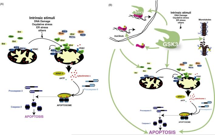
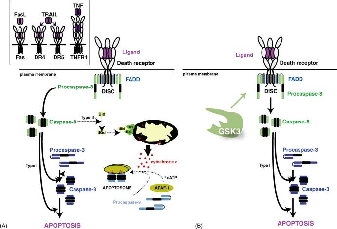
Few things can be considered to be more important to a cell than its threshold for apoptotic cell death, which can be modulated up or down, but rarely in both directions, by a single enzyme. Therefore, it came as quite a surprise to find that one enzyme, glycogen synthase kinase-3 (GSK3), has the perplexing capacity to either increase or decrease the apoptotic threshold. These apparently paradoxical effects now are known to be due to GSK3 oppositely regulating the two major apoptotic signaling pathways. GSK3 promotes cell death caused by the mitochondrial intrinsic apoptotic pathway, but inhibits the death receptor-mediated extrinsic apoptotic signaling pathway. Intrinsic apoptotic signaling, activated by cell damage, is promoted by GSK3 by facilitation of signals that cause disruption of mitochondria and by regulation of transcription factors that control the expression of anti- or pro-apoptotic proteins. The extrinsic apoptotic pathway entails extracellular ligands stimulating cell-surface death receptors that initiate apoptosis by activating caspase-8, and this early step in extrinsic apoptotic signaling is inhibited by GSK3. Thus, GSK3 modulates key steps in each of the two major pathways of apoptosis, but in opposite directions. Consequently, inhibitors of GSK3 provide protection from intrinsic apoptosis signaling but potentiate extrinsic apoptosis signaling. Studies of this eccentric ability of GSK3 to oppositely influence two types of apoptotic signaling have shed light on important regulatory mechanisms in apoptosis and provide the foundation for designing the rational use of GSK3 inhibitors for therapeutic interventions.


Figures

Fig. 1
Fig. 1
Mechanisms that regulate the actions of GSK3. Four mechanisms act in concert to regulate the phosphorylation of substrates by GSK3. Substrate phosphorylation by GSK3 is limited by the activity of the priming kinase which prepares the substrate for GSK3 because GSK3 most often phosphorylates primed substrates that are prephosphorylated four residues C-terminal to the GSK3 phosphorylation site. A major mechanism for inhibiting the activity of GSK3 is via serine-phosphorylation, so activity is inhibited when serine-9 of GSK3β or serine-21 of GSK3a is phosphorylated. Conversely, the activity of GSK3 is optimal when phosphorylated on tyrosine-216 of GSK3β or tyrosine-279 of GSK3a (not shown). When the substrate is prephosphorylated and GSK3 is active, with the regulatory serine dephosphorylated, two spatial restrictions also contribute to regulating the actions of GSK3, its subcellular localization and its association with other proteins in regulatory complexes. GSK3 is considered to be largely a cytosolic enzyme, but it is also associated with, or internalized in, subcellular compartments such as the nucleus, mitochondria, and growth cones, so dynamic regulation of the subcellular localization of GSK3 can regulate its access to substrates within subcellular compartments. Besides this gross cellular distribution of GSK3, its distribution in the cell is constrained by its propensity to be associated in protein complexes which provides an important mechanism for regulating its phosphorylation of specific substrates that are colocalized in such complexes. Thus, substrate-specific regulation of phosphorylation by GSK3 is achieved by regulation of the priming kinase activity, phosphorylation of GSK3, the subcellular localization of GSK3, and assembly of GSK3 in protein complexes.
Fig. 2
Fig. 2
The intrinsic apoptotic signaling pathway. (A) Depiction of the intrinsic apoptotic signaling pathway. In response to cell damaging insults, pro-apoptotic members of the bcl-2 family (e.g., Bax, Bim) are activated and they translocate to the mitochondria to neutralize anti-apoptotic proteins (e.g., bcl-2, Mcl-1, Bcl-xL). This results in disruption of mitochondria which triggers the release of pro-apoptotic molecules from the mitochondrial intermembrane space. Released cytochrome c clusters with APAF-1 and procaspase-9 in the presence of dATP to form the apoptosome to activate caspase-9. Activated caspase-9 cleaves and activates caspase-3, triggering a caspase cascade which ultimately results in the death of the cell. (B) GSK3 promotes the intrinsic apoptotic signaling pathway be regulating transcription factors that control the expression of pro- and anti-apoptotic proteins, by promoting microtubule disruption and cell structural changes that occur during apoptosis, and by promoting disruption of mitochondria.
Fig. 3
Fig. 3
The extrinsic apoptotic signaling pathway. (A) Depiction of the extrinsic apoptotic signaling pathway. The four major apoptosis-inducing death receptors are activated when Fas ligand (FasL) activates Fas, TRAIL activates DR4 or DR5, or TNF activates TNF-R1 (left inset). The binding of the ligand to the death receptor induces trimerization of the receptor which produces a conformation that recruits FADD and procaspase-8 to the cytoplasmic tail of the receptor, altogether forming a protein complex known as the DISC. Within the DISC, caspase-8 is activated by auto-cleavage. In type I cells, sufficient active caspase-8 is generated to directly activate caspase-3 to carry out the apoptotic program. In type II cells, activation of caspase-8 leads to activation of caspase-3 indirectly through cleavage and activation of Bid, forming tBid, which activates the mitochondrial apoptotic mechanisms that are involved in the intrinsic apoptotic pathway. (B) GSK3 inhibits the extrinsic apoptotic pathway by impairing transduction of the signal from activated death receptors to the activation of caspase-8.

References

    1. Adhikary S, Eilers M. Transcriptional regulation and transformation by Myc proteins. Nat. Rev. Mol. Cell. Biol. 2005;6:635–645. - PubMed
    1. Akhtar RS, Ness JM, Roth KA. Bcl-2 family regulation of neuronal development and neurodegeneration. Biochim. Biophys. Acta. 2004;1644:189–203. - PubMed
    1. Alvarez G, Munoz-Montano JR, Satrustegui J, Avila J, Bogonez E, Diaz-Nido J. Lithium protects cultured neurons against β-amyloid-induced neurodegeneration. FEBS Lett. 1999;453:260–264. - PubMed
    1. Alvarez AR, Godoy JA, Mullendorff K, Olivares GH, Bronfman M, Inestrosa NC. Wnt-3a overcomes β-amyloid toxicity in rat hippocampal neurons. Exp. Cell Res. 2004;297:186–196. - PubMed
    1. Armstrong JS. Mitochondrial membrane permeabilization: the sine qua non for cell death. Bioessays. 2006;28:253–260. - PubMed

Publication types

MeSH terms