

Review
doi: 10.1016/j.cbpa.2006.08.013.
Epub 2006 Aug 28.
Isotope-edited IR spectroscopy for the study of membrane proteins
Affiliations
- PMID: 16935550
- PMCID: PMC7185810
- DOI: 10.1016/j.cbpa.2006.08.013
Item in Clipboard
Review
Isotope-edited IR spectroscopy for the study of membrane proteins
Curr Opin Chem Biol.
2006 Oct.
Abstract
Fourier transform infrared (FTIR) spectroscopy has long been a powerful tool for structural analysis of membrane proteins. However, because of difficulties in resolving contributions from individual residues, most of the derived measurements tend to yield average properties for the system under study. Isotope editing, through its ability to resolve individual vibrations, establishes FTIR as a method that is capable of yielding accurate structural data on individual sites in a protein.
Figures

FTIR spectra of the influenza A M2 transmembrane domain (25 amino acids) reconstituted in lipid bilayers [23]. The spectra shown are of samples with no label (gray line), a single 13C 16O label (dashed line) or a single 13C18O label (black line). The location of the unlabeled and labeled peaks are shown.
16O label (dashed line) or a single 13C18O label (black line). The location of the unlabeled and labeled peaks are shown.
Schematic representation of the effect of helix geometry upon the dichroisms of three arbitrary vibrations shown in blue, yellow and green. In the left model, the three different vibration transition dipole moments are all equally aligned with respect to the z axis and hence exhibit the same dichroism. However, tilting the helix by the angle β in the middle structure now results in the green mode having the highest dichroism because it is the least tilted with respect to the z axis. Finally, rotation about the helix director by the angle ω reverses the dichroism ratio, causing the blue mode to exhibit the highest dichroism.

Structure of the bond predicted to take place in the transmembrane domain of human glycophorin A [31] and measured experimentally using isotope-edited FTIR [30••].
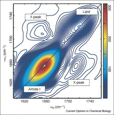
2D IR spectrum of the transmembrane helical bundle formed by the T-cell receptor CD3-ζ component reconstituted in lipid bilayers. The spectrum exhibits cross peaks between the amide I band of the protein and the lipid head-group ester carbonyl, providing proof that peptide backbone and membrane head-groups are strongly coupled.

FTIR spectra of the transmembrane domain of SARS coronavirus E protein reconstituted in phospholipid bilayers [47] in the amide I and amide II regions. The spectra were obtained after flushing air saturated with H2O (solid line) or D2O (dotted line) over the sample. Both spectra were normalized so as to adjust the absorption of the amide I mode (1658 cm−1) to 1.0 OD. Note the reduction that takes place in the amide II mode (1545 cm−1) upon D2O exchange.
References
-
- Krimm S., Bandekar J. Vibrational spectroscopy and conformation of peptides, polypeptides, and proteins. Adv Protein Chem. 1986;38:181–364. - PubMed
-
- Braiman M.S., Rothschild K.J. Fourier transform infrared techniques for probing membrane protein structure. Annu Rev Biophys Biophys Chem. 1988;17:541–570. - PubMed
-
- Surewicz W.K., Mantsch H.H., Chapman D. Determination of protein secondary structure by Fourier transform infrared spectroscopy: a critical assessment. Biochemistry. 1993;32:389–394. - PubMed
-
- Tatulian S.A. Attenuated total reflection Fourier transform infrared spectroscopy: a method of choice for studying membrane proteins and lipids. Biochemistry. 2003;42:11898–11907. - PubMed
-
- Wallace B.A., Teeters C.L. Differential absorption flattening optical effects are significant in the circular dichroism spectra of large membrane fragments. Biochemistry. 1987;26:65–70. - PubMed
Publication types
MeSH terms
Substances
Grants and funding
LinkOut - more resources
Full Text Sources
