Review
. 2006;34(15):4216-24.
doi: 10.1093/nar/gkl508. Epub 2006 Aug 25.

Mechanisms of a ring shaped helicase

Affiliations
Review

Mechanisms of a ring shaped helicase

Ilker Donmez et al. Nucleic Acids Res. 2006.

Abstract

Bacteriophage T7 helicase (T7 gene 4 helicase-primase) is a prototypical member of the ring-shaped family of helicases, whose structure and biochemical mechanisms have been studied in detail. T7 helicase assembles into a homohexameric ring that binds single-stranded DNA in its central channel. Using RecA-type nucleotide binding and sensing motifs, T7 helicase binds and hydrolyzes several NTPs, among which dTTP supports optimal protein assembly, DNA binding and unwinding activities. During translocation along single stranded DNA, the subunits of the ring go through dTTP hydrolysis cycles one at a time, and this probably occurs also during DNA unwinding. Interestingly, the unwinding speed of T7 helicase is an order of magnitude slower than its translocation rate along single stranded DNA. The slow unwinding rate is greatly stimulated when DNA synthesis by T7 DNA polymerase is coupled to DNA unwinding. Using the T7 helicase as an example, we highlight critical findings and discuss possible mechanisms of helicase action.


Figures

Figure 1
Figure 1
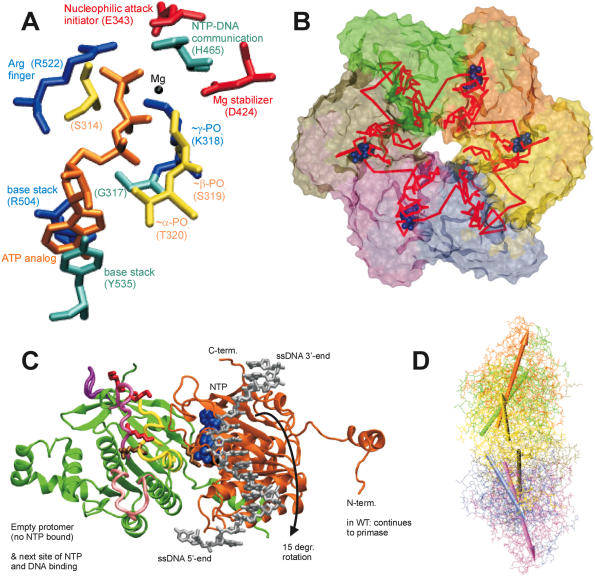
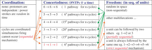
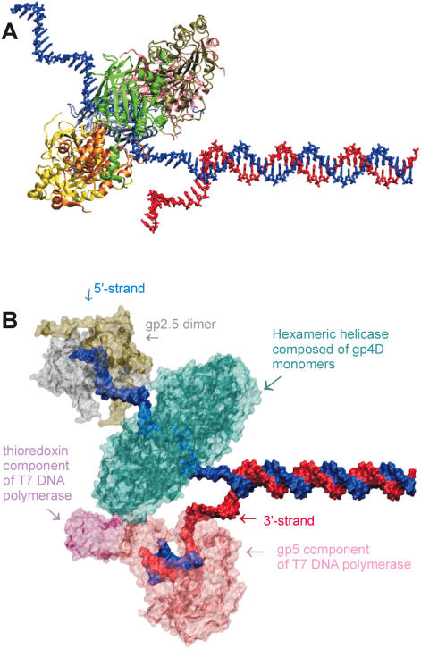
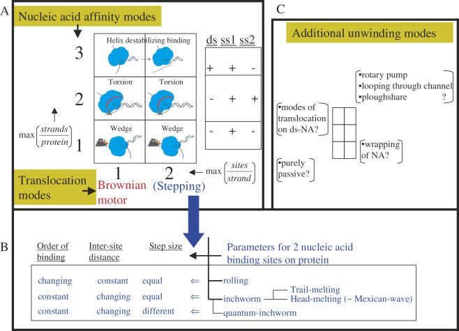
(A) Critical residues coordinating the bound nucleotide. Amino acids around the nucleotide binding site of T7 helicase are shown with brief notes on their possible functions. The nucleotide is in orange, Mg(II) in black, Arg and Lys in blue, Asp and Glu in red, Ser and Thr in yellow, His, Tyr and Gly in cyan. Among the included residues, 314, 317, 318, 319 and 320 are a part of the Walker A loop, whereas residue 424 is a part of the Walker B motif. All the residues are from the B chain in the strucutre 1e0j (17), except R522 which is from the C chain. (B) Conformational change transmission network in the structural model of the ring-shaped hexameric T7 helicase. All six subunits of the T7 helicase domain are illustrated by a surface representation and the red lines show one of the possible conformational change transmission paths with an undefined (and un-implied) directionality and sequence. The red network simply traces the inter-subunit sensor R522, the hypothetical DNA binding loops (424–439, 464–475, 503–513), the residue that is likely to establish intra-domain nucleotide ligation state communication (H465), a residue proposed to initiate the nucleophilic attack (E343), residues that disrupt helicase functioning when mutated (S345, E348, S496, R487, G488, S345, G451) (27) and eight other amino acids that are critical in coordinating or stabilizing the bound cofactor (S314, G317, K318, S319, T320, D424, R504, Y535) (17). The order of residues is as follows: 522, 451, 496, 488, 487, (475–464), (439–424), (503–413), 314, (317–320), 343, 345, 348, 535, next522. Coloring of the subunits A–F is as follows: yellow, orange, green, tan, mauve, iceblue. (C) Two helicase protomers with a possible delineation of single stranded DNA. Two subunits of the hexameric helicase are viewed from the central channel, their C-termini facing upward. The DNA is oriented with its 5′ end facing down in the figure. DNA is initially bound to the right subunit, which is bound to NTP and thus is in a high affinity state for DNA. The difference in orientations between unbound and NTP docked conformations is a rotation of 15°, which might correspond to the power stroke (17). The directionality of the rotation upon NTP docking (shown with an arrow) agrees with the proposed orientation of DNA. The probability of DNA being caught by the left subunit that supplies the Arginine finger is increased both because of its likelihood of binding to NTP in the next step and because of its vicinity to the center of the ring [5 å closer than the other neighbor (17)]. We took the single DNA strand from NDB code bd0070 and manually positioned it on a dimer from PDB code 1e0j (17). We used VMD and its associated routines (62,63) for visualization and POV-Ray () for rendering. The ligand-free subunit is in green (chain C in the structure), the NTP bound one is in orange (chain B). The DNA is shown in silver and the nucleotide in blue. The putative DNA binding loops [424–439 in pink (lowermost), 464–475 in mauve (middle), 503–513 in purple (uppermost loop)] (17), the motif H4 (480–500 in yellow) and the mutationally tested residues on them are also indicated (K467, K471, K473 in red R487, R522 in tan) on the C subunit (green subunit). (D) Relative orientations of helicase monomers. To get an idea for the inherent symmetry in a hexameric ring, we can look at the orientations of the six monomers with respect to a common reference, such as the line passing through the central channel through bound DNA. If for all subunits, a chosen line within the monomer that connects one of its residues close to the inner channel to another one distal to the inner channel, is equally perpendicular to the line of DNA, then we would conclude that the monomers are symmetrically arranged with respect to the above chosen definition. When we apply this type of an analysis to T7 hexamer (17), we see that the monomers are not arranged symmetrically (17). For instance the upper part of the figure has A, B and C subunits in yellow, orange and green. A is closest to the screen and C is the distal one. As seen, there is an apparent rotation (∼15°) between successive units (17). To relate to the mechanism shown in (C) [the DNA in (D) has its 5′ end towards the right side of the figure], we can use two simple approaches: As the DNA is initially bound to subunit B, upon NTP hydrolysis the orientation of subunit B would become similar to that of subunit A (because the electron density in the crystal structure was lesser for subunit A nucleotide than for subunit B nucleotide, thus A is more like an NDP state whereas B is more like an NTP state) (17), as a result the B subunit rotation would be in a direction to extrude DNA towards the 5′end (right side). As a second approach to interpret the figure, we can consider the biochemical finding that the NTP docking is the power stroke step (32): in this case after an empty subunit (such as C) receives DNA, it would move it as soon as the nucleotide docks and as the subunit C is already in the maximally rotated state, the only way it can rotate will be in the direction to reset its orientation to that of monomer B (since C is binding NTP as it is doing this, it is already natural for it to assume the state of the NTP bound monomer). In both cases the direction of extruding DNA is toward the right, ie. the 5′ end of DNA, thus making the helicase movement 5′–3′. The relative arrangement of the upper trimer and the lower trimer is not significant, as the lower one was just modeled upon the upper one via symmetry assumptions. The coloring is the same as in (B), arrows having the same colors as the monomers that they represent.
Figure 2
Figure 2
Possible NTPase mechanisms for ring-shaped helicases. Three factors define the nature of the NTPase mechanism employed by the ring subunits (32) and these are arranged in the three columns. (i) Coordination defines the deterministic nature of subunit firing: If firings are random in time, a single firing event by a subunit can be followed by co-firing by two subunits or more, thus there is no regulation on the number of units that hydrolyze NTP and employ the power stroke to the DNA at the same time. On the other hand, if the cycles are fully coordinated, a firing unit of a certain number of protomers has to wait until the preceding unit of the same size finishes firing. (ii) Concertedness or the number of subunits that hydrolyze NTP at a time (all-concerted versus paired versus 1 by 1): This is simply the number of subunits that hydrolyze NTP at the same time. (iii) Freedom or the degeneracy of permutations in the firing sequence. A firing by a certain number of subunits is followed by firing by any other similarly sized unit if firings are random in space, thus firing by first unit can be followed by any of the available units of the same size. On the contrary, if the firings are non-random, they would have a defined sequentiality: a certain unit will be more likely to be followed by another certain unit. The simplest mechanism that agrees with the data on T7 helicase is a partially or strictly sequential mode with a firing unit size of one monomer (32).
Figure 3
Figure 3
(A) Strand exclusion model: T7 helicase ring (4D domain) positioned on the 5′ strand of DNA. An illustration of the wedge mechanism of unwinding: one of the strands of DNA is threaded through the central channel of the ring while the other one is left outside. (B) Components of the T7 replisome assembled on fork DNA. A possible assembly of T7 DNA polymerase (gp5 complexed with Escherichia coli thioredoxin), T7 single stranded binding protein (gp2.5, active as a dimer), and T7 helicase 4D domains as a hexameric ring. The excluded molecules are the primase domain of helicase and the second DNA polymerase on the lagging strand. We used X3DNA (64) to model DNA structure of a random DNA sequence, Swiss PDB Viewer (65) for manually rearranging and docking of protein and DNA structures, VMD and its associated routines (62,63) for representations and POV-Ray () for rendering the structures. DNAP is from 1t7p (66), gp2.5 is from 1je5 (67) and helicase the gp4D fragments from 1e0j (17). DNAP is in pink/mauve, gp2.5 dimer in silver/tan, gp4D hexamer in cyan, 5′ strand in blue and 3′ strand in red.
Figure 4
Figure 4
Possible mechanisms of helicase translocation and nucleic acid unwinding. Nucleic acid unwinding is considered to be a combination of two processes: unidirectional translocation (achieved by the protein via polar binding to DNA) and strand separation (a function of transient sets of nucleic acid affinity modes of the protein). The six-part box in (A) illustrates the possible unwinding modes resulting from the various nucleic acid binding modes (y-axis) and translocation modes (x-axis). The second box on the right in (A) shows an experimental scheme that parallels the three nucleic acid binding modes of the y-axis. For example, if the only type of binding between the protein and DNA is one protein site bound to one strand of the DNA, then the stoichiometry max (strands/protein) would be 1. If the maximal stoichiometry is two strands per protein the value would be 2, and if one protein can bind to both duplex and single stranded nucleic acid, the y-axis value would be 3 (= 2 + 1). The nucleic acid affinity modes outlined on the y-axis define the unwinding mechanisms depicted with pictures within the boxes. The wedge unwinding mode results from the helicase interacting exclusively with one strand, a torsional component of unwinding would necessitate interaction with an additional strand of nucleic acid, and direct destabilization of the nucleic acid junction would require interaction with the duplex region at the junction in addition to the single strand on which the helicase is translocating. The x-axis defines the translocation mechanisms, and the two numerical values it can take correspond to the maximum observable number of nucleic acid sites that the helicase can transiently and simultaneously bind to during translocation. If there is only one possible nucleic acid binding site, the helicase may move unidirectionally along the nucleic acid using a Brownian motor (BM) or ratchet type mechanism (68,69). If the helicase binds the nucleic acid via two sites, it can employ stepping modes of translocation, such as rolling or inchworm. The stepping translocation modes are further elaborated in (B). In the rolling mechanism, the two helicase sites that bind successive regions of the nucleic acid alternate their affinity while retaining the distance between them but not their relative order. In other words, one helicase site is ahead in one step, but the other one moves in front during the next step. In the inchworm mechanism, the helicase polarity is maintained during translocation; thus, the same side of the protein faces the same end (3′ or 5′) of the nucleic acid at all times. To move, the distance between the two protein sites and the binding affinity periodically changes. A widely accepted mechanism combines the inchworm translocation mode with the duplex destabilization mode. In this mechanism, the two contact sites, protein–dsDNA and protein–ssDNA, can be in one of two relative orders. If the dsDNA binding site is ahead of the other, the mechanism might be referred to as head-melting, or trail-melting in the other case. The mode of head-melting can be likened to a Mexican-wave movement along the DNA (70,71). In the case where these two sites take steps of different sizes, the mechanism was given the name quantum inchworm (72). Further modifications of inchworm mechanism taking into account requirements for the nature of the displaced strand or the predominant mode of interaction with the loading strand (i.e. hydrophobic or electrostatic) have also been proposed (–75). We display all six combinations as physically plausible, even though not all have been proposed previously. The most popular mechanisms are shown by the coordinates (1,1), (1,2) and (2,3).In addition to the above modes, several other types of unwinding mechanisms have been proposed as illustrated in (C). For example a purely passive mechanism is probably employed by the chemical formaldehyde (76), ssRNA wrapping might be used by Rho, dsDNA translocation might be used by DnaB during branch migration (15), ploughshare is proposed for MCM proteins (77), and looping through channel is proposed for SV40-large T antigen (78). In addition, the torsional model can work with a duplex DNA in the central channel in addition to the mentioned mode of binding two single strands separately. Classifying most of these specific mechanisms requires extra dimensions in the 2 × 3 graph of part (A).

Similar articles

Cited by

References

    1. Matson S.W., Bean D.W., George J.W. DNA helicases: enzymes with essential roles in all aspects of DNA metabolism. BioEssays. 1994;16:13–22. - PubMed
    1. Lohman T.M., Bjornson K.P. Mechanisms of helicase-catalyzed DNA unwinding. Annu. Rev. Biochem. 1996;65:169–214. - PubMed
    1. Patel S.S., Picha K.M. Structure and function of hexameric helicases. Annu. Rev. Biochem. 2000;69:651–697. - PubMed
    1. van Brabant A.J., Stan R., Ellis N.A. DNA helicases, genomic instability, and human genetic disease. Annu. Rev. Genomics Hum. Genet. 2000;1:409–459. - PubMed
    1. Mohaghegh P., Hickson I.D. DNA helicase deficiencies associated with cancer predisposition and premature ageing disorders. Hum. Mol. Genet. 2001;10:741–746. - PubMed

Publication types

MeSH terms