. 2006 Nov 15;91(10):3684-91.
doi: 10.1529/biophysj.106.088062. Epub 2006 Aug 25.

Mechanosensitive channel gating transitions resolved by functional changes upon pore modification

Affiliations

Mechanosensitive channel gating transitions resolved by functional changes upon pore modification

Jessica L Bartlett et al. Biophys J. .

Abstract

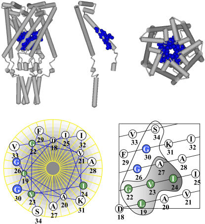
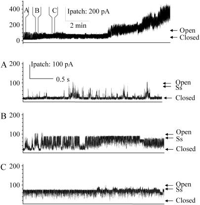
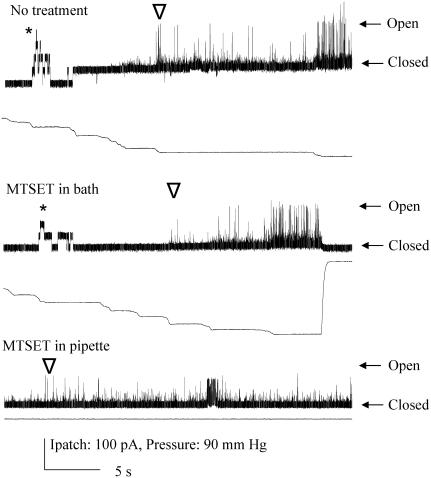
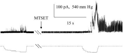
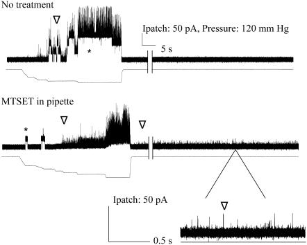
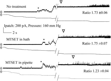
The mechanosensitive channel of large conductance acts as a biological "emergency release valve" that protects bacterial cells from hypoosmotic stress. Although structural and functional studies and molecular dynamic simulations of this channel have led to several models for the structural transitions that occur in the gating process, inconsistencies linger and details are lacking. A previous study, using a method coined as the "in vivo SCAM", identified several residues in the channel pore that were exposed to the aqueous environment in the closed and opening conformations. Briefly, the sulfhydryl reagent MTSET was allowed to react, in the presence or absence of hypoosmotic shock, with cells expressing mechanosensitive channel of large conductance channels that contained cysteine substitutions; channel dysfunction was assessed solely by cell viability. Here we evaluate the MTSET-induced functional modifications to these mechanosensitive channel activities by measuring single channel recordings. The observed changes in residue availability in different states, as well as channel kinetics and sensitivity, have allowed us to elucidate the microenvironment encountered for a number of pore residues, thus testing many aspects of previous models and giving a higher resolution of the pore domain and the structural transitions it undergoes from the closed to open state.


Figures

FIGURE 1
FIGURE 1
Schematic depictions of the E. coli MscL emphasizing the pore domain and specific residues that were targeted for substitutions. The upper panel shows a model for the closed MscL structure (11) based upon the crystal structure (10). The residues investigated by this study are highlighted in blue and shown in a cpk format. A side view (left), a single subunit of the pentameric complex (center), and top view are shown. The bottom panel presents an idealized helical wheel (left) and net (right) of the E. coli MscL first transmembrane domain. The residues encircled in the helical net were identified in the in vivo SCAM assay (17) as described in text. The residues within the shaded region were accessible to MTSET only upon channel gating by osmotic downshock. The residues that are further investigated by patch clamp in this study are colored dependent on whether the MTSET was accessible without (blue) or with (green) channel gating.
FIGURE 2
FIGURE 2
G26C locks in an open channel conformation when modified by MTSET placed on the periplasmic side. The uppermost trace shows G26C activity with MTSET added by backfilling the pipette as described in Materials and Methods, note that no pressure was applied before or during this trace. At the time points indicated by panels A, B, and C, the trace has been expanded. Panel A shows the first channel starting to open and the preference for substates and short open dwell times. Panel B shows the first channel being locked into an open state. Panel C shows the final preference of the channel for a common 4/5ths open substate (labeled as Ss).
FIGURE 3
FIGURE 3
G30C shows gating-independent spontaneous activity when modified by MTSET placed on the periplasmic, but not bath cytoplasmic side. Single channel recordings of G30C are shown from top to bottom without treatment, with MTSET added to the bath, and with MTSET added to the periplasmic side of the patch by backfill, as described in text. Below each channel trace, the amount of pressure stimulation is shown. Note that in the top two traces, MscS activity (indicated by *) is seen before MscL activity (indicated by ∇). The arrows indicate the closed and the normal highest conducting open state for the MscL channels shown. In the final trace, G30C shows spontaneous gating with very short open dwell times; full openings are not often resolved.
FIGURE 4
FIGURE 4
G26C is accessible from the cytoplasmic side only subsequent to channel gating. Single channel recordings of G26C were observed upon normal pressure stimulation (left; below the channel trace the amount of pressure stimulation is shown). After addition of MTSET to the bath (as indicated at arrow), no channel activity is observed, even after tens of seconds (middle of trace). Once channel gating is effected by pressure, channels are observed to gate spontaneously (right portion of trace).
FIGURE 5
FIGURE 5
V23C shows gating-dependent spontaneous activity when modified by MTSET placed on the periplasmic, but not cytoplasmic side. Channel traces of untreated (top) and treated (bottom) patches containing native membranes expressing V23A. In the bottom trace, treatment was effected by filling the pipette with MTSET-containing buffer. No channel activity is initially observed (left part of both traces), but can be induced by suction in the pipette (pressure is shown below each channel trace). In both traces MscS activity (indicated by *) and MscL activity (indicated by ∇) are observed (MscL rides up upon the MscS activity in the top trace). Note that in the bottom trace, subsequent to pressure-induced gating, a “flickery” channel with extremely short open dwell times is observed; this is shown in the bottom trace, which is a blowup of the indicated region. Hash marks represent ∼3–5 min removed to show durability of response.
FIGURE 6
FIGURE 6
I24C shows gating-independent increased sensitivity when modified by MTSET on the periplasmic, but not cytoplasmic side. Single channel recordings of I24C illustrate the change in pressure sensitivity upon addition of MTSET on the periplasmic side. The “ratio”, which is the average mean ± SE from multiple patches, is derived from the relative pressures for opening MscL/MscS, as described in Materials and Methods; a larger ratio indicates a channel that requires more tension to open. Both the MscS (*) and MscL (∇) activities are shown. The first or topmost trace is I24C with no treatment. The second trace is the same patch with MTSET added to the cytoplasm and it has been exercised multiple times. The third trace is obtained where the pipette was backfilled with MTSET as described in Materials and Methods. Hatch marks indicate 20–30 s.

References

    1. Kloda, A., and B. Martinac. 2001. Mechanosensitive channels in archaea. Cell Biochem. Biophys. 34:349–381. - PubMed
    1. Sukharev, S., and D. P. Corey. 2004. Mechanosensitive channels: multiplicity of families and gating paradigms. Sci. STKE. 2004:re4. - PubMed
    1. Sukharev, S. I., P. Blount, B. Martinac, F. R. Blattner, and C. Kung. 1994. A large-conductance mechanosensitive channel in E. coli encoded by MscL alone. Nature. 368:265–268. - PubMed
    1. Moe, P., and P. Blount. 2005. Assessment of potential stimuli for mechano-dependent gating of MscL: effects of pressure, tension, and lipid headgroups. Biochemistry. 44:12239–12244. - PubMed
    1. Levina, N., S. Totemeyer, N. R. Stokes, P. Louis, M. A. Jones, and I. R. Booth. 1999. Protection of Escherichia coli cells against extreme turgor by activation of MscS and MscL mechanosensitive channels: identification of genes required for MscS activity. EMBO J. 18:1730–1737. - PMC - PubMed

Publication types

MeSH terms

LinkOut - more resources