

Analysis of the mononuclear inflammatory cell infiltrate in the normal breast, benign proliferative breast disease, in situ and infiltrating ductal breast carcinomas: preliminary observations
- PMID: 16935972
- PMCID: PMC1860493
- DOI: 10.1136/jcp.2005.031252
Analysis of the mononuclear inflammatory cell infiltrate in the normal breast, benign proliferative breast disease, in situ and infiltrating ductal breast carcinomas: preliminary observations
Abstract
Background: Mammary carcinogenesis is a multistep process entailing the transition from normal breast to benign proliferative breast disease (ductal hyperplasia) to ductal carcinoma in situ to infiltrating ductal carcinoma.
Hypothesis: These transitions are associated with changes in the mononuclear inflammatory cell infiltrate.
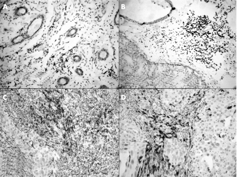
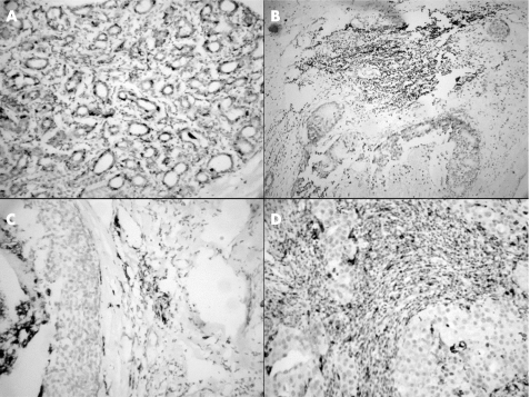
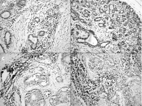
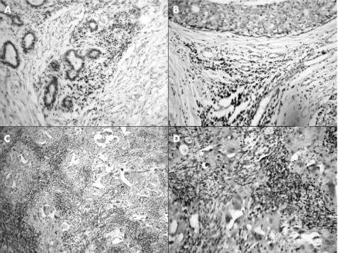
Materials and methods: A total of 53 mastectomy specimens of normal breast, benign proliferative breast disease, ductal carcinoma in situ and infiltrating ductal carcinoma were evaluated for mononuclear inflammatory cell infiltrate by using immunohistological methods and monoclonal antibodies including CD20, CD68, CD3 and granzyme B, histiocytes, T cells and cytotoxic T cells.
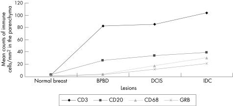
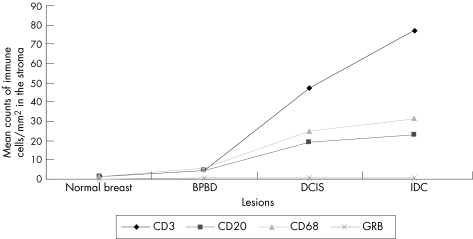
Results: Transitions from normal breast to the subsequent tissue with lesions (normal skin v benign proliferative breast disease v ductal carcinoma in situ v infiltrating ductal carcinoma) were associated with significantly (p<0.01) increased mean (SD) density of mononuclear inflammatory cell infiltrate at the parenchyma (3.2 (1.0) v 26.4 (7.8) v 33.6 (7.9) v 39.1 (4.7) for CD20+ B cells; 2.8 (1.0) v 81.5 (14.0) v 84.0 (14.9) v103.7 (3.9) for CD3; 1.3 (2.0) v 3.8 (4.0) v 12.7 (23) v 22.1 (25.0) for CD68+ macrophages; 2.0 (1.0) v 58.3 (5.0) v 60.0 (10.0) v 74.1 (28.0) for granzyme B+ cytotoxic T cells) and at the stroma (0.7 (1.0) v 3.0 (5.0) v 13.3 (20) v 16.7 (30.0) for CD20+ B cells; 1.0 (2.06) v 4.0 (2.5) v 16.7 (5.0) v 21.7 (15) for CD68+ macrophages; 1.4 (0.6) v 4.2 (1.2) v 46.6 (16.7) v 77.0 (5.0) for CD3+ cells and 0 (0) v 0.5 (1.0) v 0.7 (1.0) v 0.7 (1.0) for granzyme B+ cytotoxic T cells).
Conclusions: The increased mononuclear inflammatory cell infiltrate during mammary carcinogenesis may reflect non-specific or specific immunological processes.
Conflict of interest statement
Competing interests: None declared.
References
-
- Hussein M R, Ismael H H. Alterations of p53, Bcl‐2, and hMSH2 protein expression in the normal breast, benign proliferative breast disease, in situ and infiltrating ductal breast carcinomas in the upper Egypt. Cancer Biol Ther 20043983–988. - PubMed
-
- Alam S M, Clark J S, Leech V.et al T cell receptor gamma/delta expression on lymphocyte populations of breast cancer patients. Immunol Lett 199231279–283. - PubMed
-
- Chin Y, Janseens J, Vandepitte J.et al Phenotypic analysis of tumor‐infiltrating lymphocytes from human breast cancer. Anticancer Res 1992121463–1466. - PubMed
-
- Baxevanis C N, Dedoussis G V, Papadopoulos N G.et al Tumor specific cytolysis by tumor infiltrating lymphocytes in breast cancer. Cancer 1994741275–1282. - PubMed
-
- Marsigliante S, Biscozzo L, Marra A.et al Computerised counting of tumour infiltrating lymphocytes in 90 breast cancer specimens. Cancer Lett 199913933–41. - PubMed
MeSH terms
LinkOut - more resources
Full Text Sources
Medical