. 2006 Nov;188(22):7785-95.
doi: 10.1128/JB.00673-06. Epub 2006 Aug 25.

Biofilm formation by Streptococcus pneumoniae: role of choline, extracellular DNA, and capsular polysaccharide in microbial accretion

Affiliations

Biofilm formation by Streptococcus pneumoniae: role of choline, extracellular DNA, and capsular polysaccharide in microbial accretion

Miriam Moscoso et al. J Bacteriol. 2006 Nov.

Abstract

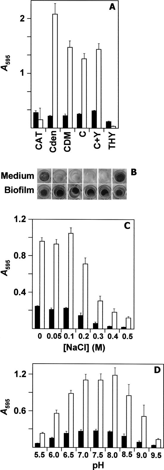
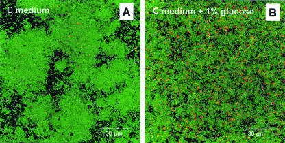
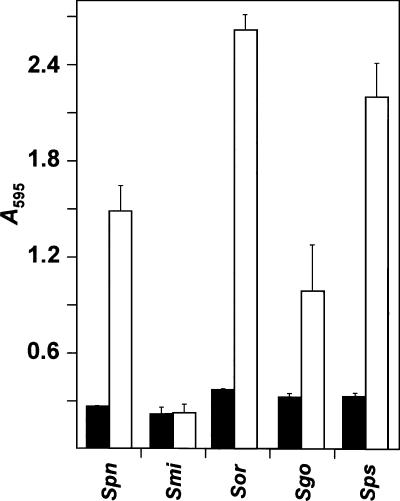
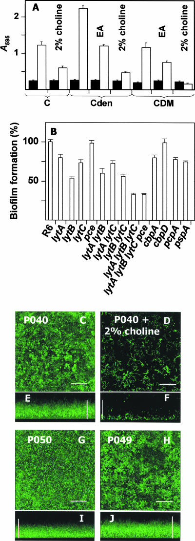
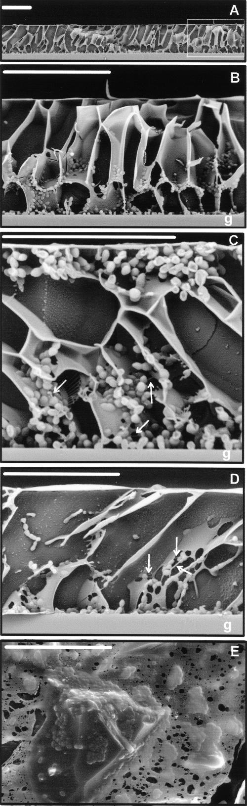
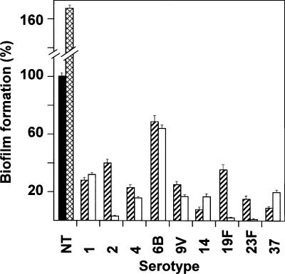
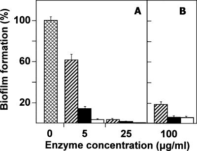
Streptococcus pneumoniae colonizes the human upper respiratory tract, and this asymptomatic colonization is known to precede pneumococcal disease. In this report, chemically defined and semisynthetic media were used to identify the initial steps of biofilm formation by pneumococcus during growth on abiotic surfaces such as polystyrene or glass. Unencapsulated pneumococci adhered to abiotic surfaces and formed a three-dimensional structure about 25 microm deep, as observed by confocal laser scanning microscopy and low-temperature scanning electron microscopy. Choline residues of cell wall teichoic acids were found to play a fundamental role in pneumococcal biofilm development. The role in biofilm formation of choline-binding proteins, which anchor to the teichoic acids of the cell envelope, was determined using unambiguously characterized mutants. The results showed that LytA amidase, LytC lysozyme, LytB glucosaminidase, CbpA adhesin, PcpA putative adhesin, and PspA (pneumococcal surface protein A) mutants had a decreased capacity to form biofilms, whereas no such reduction was observed in Pce phosphocholinesterase or CbpD putative amidase mutants. Moreover, encapsulated, clinical pneumococcal isolates were impaired in their capacity to form biofilms. In addition, a role for extracellular DNA and proteins in the establishment of S. pneumoniae biofilms was demonstrated. Taken together, these observations provide information on conditions that favor the sessile mode of growth by S. pneumoniae. The experimental approach described here should facilitate the study of bacterial genes that are required for biofilm formation. Those results, in turn, may provide insight into strategies to prevent pneumococcal colonization of its human host.


Figures

FIG. 1.
FIG. 1.
Some factors influencing biofilm formation in S. pneumoniae R6 strain. (A) Effect of the culture medium. (B) Biofilm formation by S. pneumoniae on PST microtiter plates in different media for 6 h at 34°C. Cells were stained with CV and the plates were washed to remove unattached cells. As a control, wells with medium but without bacteria were also stained with CV. The results of a representative experiment are shown. (C) Effect of ionic strength. (D) The influence of the initial pH of the medium. In all cases, filled and open bars indicate bacterial growth and biofilm formation, respectively. The data represent the average of six samples. Standard error bars are shown.
FIG. 2.
FIG. 2.
CLSM image of the viability of biofilm-grown S. pneumoniae as a function of the incubation time. S. pneumoniae R6 cells were incubated in C medium to an A595 value of 0.5. The culture was centrifuged, and the cells were resuspended in an equal volume of prewarmed C medium. A portion of the culture was diluted 1/1,000 in the indicated medium. One percent glucose stimulates cell growth and the pH of the medium drops to below 6.0; this pH is nonpermissive for autolytic activity. After incubation at 34°C for 12 h (A) or 24 h (B), the cells in the biofilms were stained with the BacLight kit showing living (green fluorescence) and dead (red fluorescence) bacteria. Images are horizontal 3D reconstructions of 25 scans in the x-y plane.
FIG. 3.
FIG. 3.
Biofilm formation capacity of various streptococcal species of the mitis group. Smi, Sor, Sgo, and Sps indicate, respectively, the type strain of S. mitis, S. oralis, S. gordonii, and S. pseudopneumoniae. The S. pneumoniae R6 strain (Spn) was used as a control. Cells were grown in C medium for 6 h at 34°C. Filled and open bars indicate growth and biofilm formation, respectively. The results are the average of three independent experiments.
FIG. 4.
FIG. 4.
Influence of choline and CBPs on biofilm formation and CSLMs of S. pneumoniae biofilms. (A) Influence of choline and EA on biofilm formation by the R6 strain. Filled and open bars indicate growth and biofilm formation, respectively. (B) Biofilm formation capacity of several mutant strains. In this experiment, the following strains were incubated in C medium for 6 h: R6, M32 (lytA), R6B (lytB), R6C (lytC), R6D (pce), M31B (lytA lytB), P042 (lytA lytC), R6BC (lytB lytC), M31BC (lytA lytB lytC), M31BCD (lytA lytB lytC pce), P064 (cbpA), R1582 (cbpD), P065 (pcpA), and P066 (pspA). The values of biofilm formation were normalized for absorbance, and the percentages were calculated in relation to strain R6. Shown are CLSM images of pneumococcal strains harboring pMV158GFP: P040 (R6), P049 (pce), and P050 (lytA lytC). (C, D, G, and H) Horizontal 3D reconstructions of 55 scans in the x-y plane. (E, F, I, and J) Vertical 3D reconstructions of 65 scans in the x-z plane. In all of the images, the bar corresponds to 30 μm.
FIG. 5.
FIG. 5.
LTSEM of S. pneumoniae R6 biofilm. (A) General view of the biofilm formed on the surface of a glass coverslip. (B) Magnification of the area indicated by a rectangle in panel A. (C and D) Two different views. Filamentous material (indicated by arrows) links pneumococcal cells to each other and to the intercellular matrix. (E) Apical view of the irregular surface of a pneumococcal biofilm. Microcolonies of different sizes can be seen. In all micrographs, the bar indicates 20 μm.
FIG. 6.
FIG. 6.
Biofilm formation by encapsulated S. pneumoniae strains. Clinical pneumococcal isolates of the indicated serotypes were grown in C medium for 6 h at 34°C. Biofilm formation (hatched bars) was quantified by staining with CV. Open bars correspond to biofilms developed by encapsulated transformants of the indicated serotype using the unencapsulated M11 strain as recipient. NT corresponds to the M11 strain, which was used as control (filled bar) and to the nontypeable pneumococcal isolate ST344 (cross-hatched bar). The values of biofilm formation by encapsulated strains were normalized for absorbance, and the percentage was presented and normalized for the control strain M11. The percentages shown are the average of three independent experiments.
FIG. 7.
FIG. 7.
Inhibition of biofilm development in S. pneumoniae cultures in the presence of DNase or proteases. (A) S. pneumoniae R6 was grown overnight at 37°C to an A595 value of 0.5 to 0.6 in C+Y medium, centrifuged, and adjusted to an A595 of 0.6 with fresh medium. Afterwards, the cell suspension was diluted 10-fold, and 200-μl aliquots were distributed in the wells of a microtiter plate, which was then incubated for 6 h at 34°C (cross-hatched bars). Other samples received either DNase I (hatched bars), trypsin (filled bars), or proteinase K (open bars) at the indicated concentrations and were incubated as above. (B) After biofilm development (6 h at 34°C), DNase I (hatched bars), trypsin (filled bars), or proteinase K (open bars) was added at 100 μg ml−1, and incubation proceeded for an additional 1 h at 34°C before staining with CV to quantify biofilm formation.

References

    1. Akerley, B. J., E. J. Rubin, A. Camilli, D. J. Lampe, H. M. Robertson, and J. J. Mekalanos. 1998. Systematic identification of essential genes by in vitro mariner mutagenesis. Proc. Natl. Acad. Sci. USA 95:8927-8932. - PMC - PubMed
    1. Allegrucci, M., F. Z. Hu, K. Shen, J. Hayes, G. D. Ehrlich, J. C. Post, and K. Sauer. 2006. Phenotypic characterization of Streptococcus pneumoniae biofilm development. J. Bacteriol. 188:2325-2335. - PMC - PubMed
    1. Austrian, R. 1986. Some aspects of the pneumococcal carrier state. J. Antimicrob. Chemother. 18 (Suppl. A):35-45. - PubMed
    1. Berrón, S., A. Fenoll, M. Ortega, N. Arellano, and J. Casal. 2005. Analysis of the genetic structure of nontypeable pneumococcal strains isolated from conjunctiva. J. Clin. Microbiol. 43:1694-1698. - PMC - PubMed
    1. Bogaert, D., R. de Groot, and P. W. M. Hermans. 2004. Streptococcus pneumoniae colonization: the key to pneumococcal disease. Lancet Infect. Dis. 4:144-154. - PubMed

Publication types

MeSH terms

LinkOut - more resources