. 2006 Sep;169(3):943-53.
doi: 10.2353/ajpath.2006.051319.

The Schistosoma mansoni hepatic egg granuloma provides a favorable microenvironment for sustained growth of Leishmania donovani

Affiliations

The Schistosoma mansoni hepatic egg granuloma provides a favorable microenvironment for sustained growth of Leishmania donovani

Mohammed F Hassan et al. Am J Pathol. 2006 Sep.

Abstract

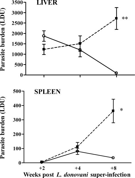
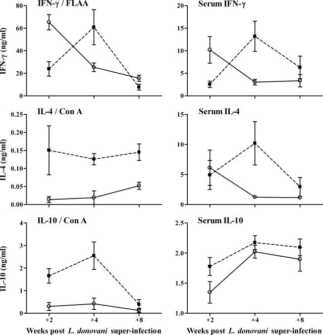
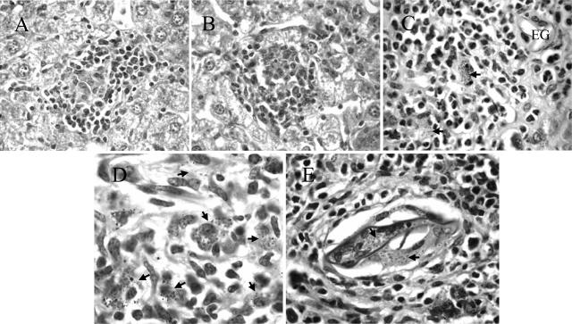
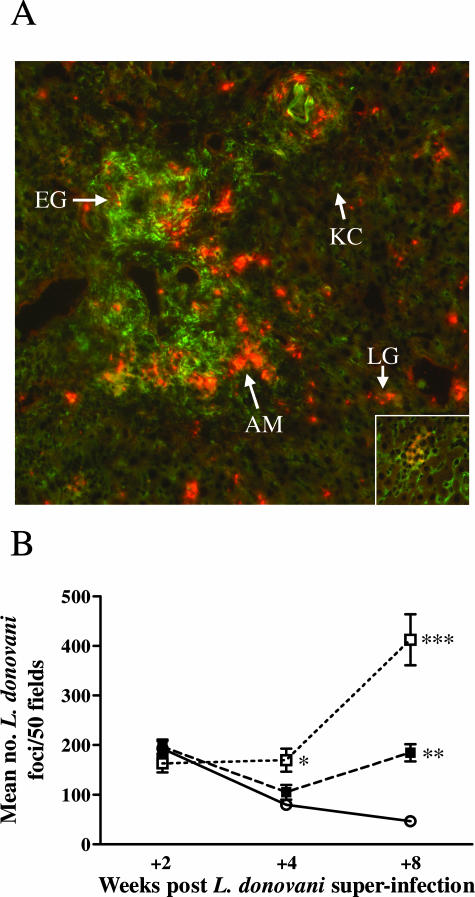
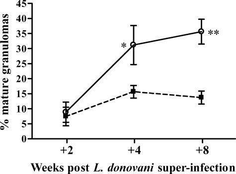
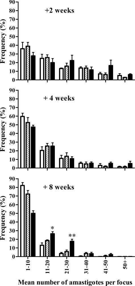
Parasitic co-infections are prevalent in many parts of the world. However, relatively little is known about how an underlying infection may impact on the host's ability to control a newly acquired parasite, especially if both infect the same organ. We have studied this using an experimental co-infection model in C57BL/6 mice involving Schistosoma mansoni and Leishmania donovani, two important human pathogens affecting the liver. We show that mice with established S. mansoni infections fail to control L. donovani growth in the liver and spleen. The failure occurs despite the development of a functional anti-L. donovani Th1 response that can mediate granuloma formation and effective clearance of amastigotes from foci of infection in the hepatic parenchyma. Instead, anti-leishmanial immunity fails within the S. mansoni egg granuloma, consistent with a lack of L. donovani granuloma assembly in this tissue microenvironment and consequent lack of NO production. Persisting amastigote replication in the S. mansoni egg granulomas may thus explain the increased L. donovani burden in the liver and spleen. These results may have implications for human S. mansoni and L. donovani co-infections and also demonstrate that granulomatous tissue responses to helminth organisms can form a discrete niche facilitating survival of intracellular pathogens.


Figures

Figure 1
Figure 1
Mice infected with S. mansoni fail to control a superimposed L. donovani infection in both the liver and the spleen. The figure shows the course of L. donovani infection in the livers and spleens of C57BL/6 mice infected with L. donovani alone (L, open circle) or infected with L. donovani at 8 weeks after infection with 25 S. mansoni cercariae (S/L, filled square). Data represent the mean ± SE Leishman-Donovan unit (LDU) values from five to six mice per group at each time point and are representative of four separate experiments. **P < 0.001, *P < 0.005.
Figure 2
Figure 2
S/L mice show elevated IL-4 and IL-10 responses and delayed development of L. donovani-specific IFN-γ responses compared to L mice. The left graphs show cytokine levels in supernatants of spleen cell cultures stimulated with ConA or formalin-fixed L. donovani amastigote antigen (FLAA). The right graphs show serum cytokine levels (mean levels in naïve mouse sera: IFN-γ, 0.393 ± 0.089; IL-4, 0.882 ± 0.017; IL-10, 1.450 ± 0.066 ng/ml). Data represent the mean ± SE cytokine levels from three mice per group for the splenocyte responses and five mice per group for the serum cytokines at each time point. FLAA-specific and serum IFN-γ levels were significantly higher in L mice (open circle) than in S/L mice (filled square) at +2 weeks (P < 0.02). Serum IFN-γ levels were significantly higher in S/L mice than in L mice at +4 weeks (P < 0.02). IL-4 and IL-10 splenocyte responses were significantly higher in S/L than in L mice (IL-4: P < 0.02 and < 0.05 at +4 and +8 weeks, respectively; IL-10: P = 0.02 and < 0.05 at +2 and +4 weeks, respectively). Serum IL-4 levels were significantly higher in SL than in L mice at +4 weeks (P < 0.05).
Figure 3
Figure 3
Morphologically normal L. donovani granulomas develop in the liver parenchyma but not in the egg granulomatous areas of S/L mice. H&E-stained sections of liver show comparable mature L. donovani granulomas in the parenchyma of L (A) and S/L (B) mice. C and D: Dispersed amastigote-laden macrophages (arrows) within the egg granulomas of S/L mice do not elicit discrete cellular foci. EG, S. mansoni egg. E: Heavy accumulation of amastigotes (arrows) within giant cells occupying a schistosome egg shell at the center of an egg granuloma. All sections are from mice at +8 weeks after superinfection. Original magnifications: ×400 (A, B); ×1000 (C, E); ×1600 (D).
Figure 4
Figure 4
Preferential generation of L. donovani foci within the schistosome egg granulomatous tissue. A: Liver sections from S/L mice at +8 weeks after co-infection were stained for L. donovani amastigotes using immune hamster anti-L. donovani serum and Alexa Fluor 546 goat anti-hamster IgG (red, arrow, AM) and for mannose receptor (MR) with anti-CD 206 and Alexa Fluor 488 goat anti-rat IgG (green). To ensure staining specificity, liver sections from the same S/L mice were stained with isotype control rat IgG2a (control for the MR staining) or normal hamster serum (control for the amastigote staining). No nonspecific staining was seen. Strong specific MR staining can be seen in the granulomatous tissue surrounding the S. mansoni eggs (arrow, EG). The parenchymal Küpffer cells stain weakly for MR (arrow, KC), and MR-positive cells are absent from the granulomas surrounding L. donovani amastigotes in the parenchyma (inset). A higher density of red-staining L. donovani amastigotes can be seen in foci of L. donovani infection in the periphery of the S. mansoni egg granulomas compared with inside the L. donovani granulomas (LG) in the parenchymal tissue. B: This was confirmed quantitatively in H&E-stained tissue sections by counting the density of L. donovani foci in the egg granulomatous tissue of S/L mice (S/L G, open square) compared with the surrounding parenchyma (S/L P, filled square) and the parenchyma of L mice (L, open circle). For each mouse 50 adjacent fields were counted at ×400 magnification. The data in B show the mean ± SE from five mice per group and is representative of two separate experiments. *P < 0.01 for S/L G versus L; **P < 0.0001 for S/L P versus L; ***P < 0.0001 for S/L G versus L, P < 0.005 for S/L G versus S/L P. Original magnifications: ×100 (A); ×400 (A, inset).
Figure 5
Figure 5
The proportion of mature parenchymal L. donovani granulomas in the liver increases progressively in L mice but not in S/L mice. Foci of infection were examined in 50 adjacent fields (×400 magnification) of H&E-stained liver tissue and recorded as L. donovani-infected Küpffer cells, immature or mature granulomas. The data shown are the mean percentages of mature granulomas ± SE from the S/L (filled square) and L (open circle) groups and is representative of two separate experiments. Percentages of mature granulomas were significantly higher in L mice than S/L mice at +4 and +8 weeks (n = 5 mice per group at each time point; *P < 0.05; **P < 0.002).
Figure 6
Figure 6
The failure of L. donovani control in S/L mice is not characterized by heavy accumulation of amastigotes within individual foci. Amastigotes, visualized by staining as described in Figure 4, were counted in individual foci and the frequencies of foci containing differing numbers of amastigotes determined. The data are based on examination of 50 adjacent fields (×1000 magnification, n = 5 mice) and is representative of two separate experiments. Data refer to L. donovani foci in the parenchyma of L mice (open bars), the parenchyma of S/L mice (shaded bars), and the egg granulomatous tissue of S/L mice (black bars). *Significantly higher than in the parenchyma of S/L mice (P < 0.01) and L mice (P < 0.02). **Significantly higher than in the parenchyma of S/L mice (P < 0.01) and L mice P < 0.001).
Figure 7
Figure 7
NOS-2 expression around foci of L. donovani replication is comparable in the liver parenchyma of L and S/L mice but inhibited within the S. mansoni egg granulomas. NOS-2-stained liver sections from L mice (A), S/L mice (B and C) 8 weeks after L. donovani infection. Long arrows: granulomas in parenchyma showing strong NOS-2 staining. Short arrows: low intensity NOS-2 staining around L. donovani foci associated with the S. mansoni egg granulomas. SG, schistosome egg granuloma showing strong NOS-2 staining around the egg. Original magnifications: ×100 (A, B); ×400 (C).

References

    1. Hoogstraal H, Heyneman D. Leishmaniais in the Sudan Republic. 30. Final epidemiological report. Am J Trop Med Hyg. 1969;18:1091–1210.
    1. el Gaddal AA. The Blue Nile Health Project: a comprehensive approach to the prevention and control of water-associated diseases in irrigated schemes of the Sudan. J Trop Med Hyg. 1985;88:47–56. - PubMed
    1. Zijlstra EE, Ali MS, el-Hassan AM, el-Toum IA, Satti M, Ghalib HW, Sondorp E, Winkler A. Kala-azar in displaced people from southern Sudan: epidemiological, clinical and therapeutic findings. Trans R Soc Trop Med Hyg. 1991;85:365–369. - PubMed
    1. Pearce EJ, C MK, Sun J, Taylor JJ, McKee AS, Cervi L. Th2 response polarization during infection with the helminth parasite Schistosoma mansoni. Immunol Rev. 2004;201:117–126. - PubMed
    1. Murray HW. Tissue granuloma structure-function in experimental visceral leishmaniasis. Int J Exp Pathol. 2001;82:249–267. - PMC - PubMed

Publication types

MeSH terms