. 2006 Oct;7(10):1019-22.
doi: 10.1038/sj.embor.7400790. Epub 2006 Aug 25.

MARCH-V is a novel mitofusin 2- and Drp1-binding protein able to change mitochondrial morphology

Affiliations

MARCH-V is a novel mitofusin 2- and Drp1-binding protein able to change mitochondrial morphology

Nobuhiro Nakamura et al. EMBO Rep. 2006 Oct.

Abstract

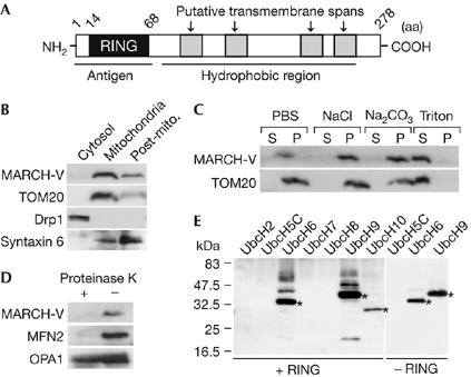
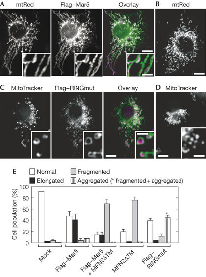
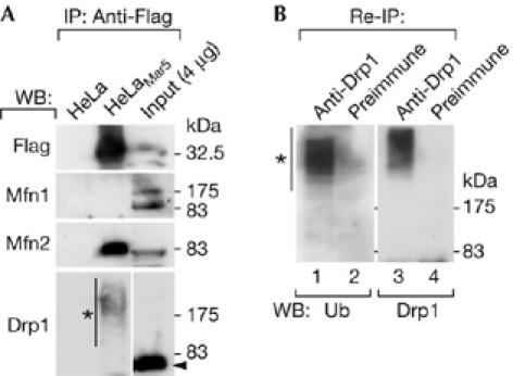
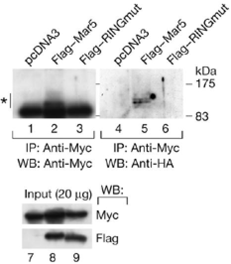
Mitofusins and Drp1 are key components in mitochondrial membrane fusion and division, but the molecular mechanism underlying the regulation of their activities remains to be clarified. Here, we identified human membrane-associated RING-CH (MARCH)-V as a novel transmembrane protein of the mitochondrial outer membrane. Immunoprecipitation studies demonstrated that MARCH-V interacts with mitofusin 2 (MFN2) and ubiquitinated forms of Drp1. Overexpression of MARCH-V promoted the formation of long tubular mitochondria in a manner that depends on MFN2 activity. By contrast, mutations in the RING finger caused fragmentation of mitochondria. We also show that MARCH-V promotes ubiquitination of Drp1. These results indicate that MARCH-V has a crucial role in the control of mitochondrial morphology by regulating MFN2 and Drp1 activities.


Figures

Figure 1
Figure 1
Characterization of MARCH-V. (A) Schematic drawing of the potential structure of MARCH-V. (B) The cytosol, mitochondria and post-mitochondria (Post-mito.) fractions (15 μg of protein) of COS7 cells were analysed by western blotting with antibodies to MARCH-V, TOM20, Drp1 and syntaxin 6 (a Golgi/endosomal protein). (C) The total membrane fractions of COS7 cells were incubated with PBS, 1 M NaCl, 0.1 M Na2CO3 (pH 11) or 1% Triton X-100 for 1 h on ice. After ultracentrifugation, the supernatant (S) and pellets (P; 20 μg of protein each) were analysed by western blotting. (D) The mitochondrial fractions were incubated with (+) or without (−) 100 μg/ml proteinase K for 1 h on ice. After quenching proteolysis by addition of phenylmethylsulphonyl fluoride (1 mM final), the mitochondria were pelleted, dissolved in SDS sample buffer and analysed by western blotting. (E) Reaction mixtures containing E1, E2 indicated above the lanes and ubiquitin (Ub) were incubated in the presence (+RING) or absence (−RING) of glutathione S-transferase fusion proteins of the MARCH-V RING finger for 2 h at 30°C. Ubiquitinated materials were detected by western blotting with anti-Ub antibody. Asterisks indicate Ub-charged E2s.
Figure 2
Figure 2
Effects of MARCH-V overexpression on mitochondrial morphology. (A,B) COS7 cells stably expressing matrix-targeted red fluorescent protein (mtRed) were transiently transfected with Flag–Mar5 and stained with Flag antibody. Signals for Flag and mtRed are shown in green and magenta, respectively. Insets show higher magnification images. A typical example of normal mitochondrial morphology in non-transfected cells is shown in (B). Scale bars, 10 (A,B) and 2 μm (A, inset). (C,D) COS7 cells transiently transfected with Flag–RINGmut were stained with Flag antibody and MitoTracker. Signals for Flag and MitoTracker are shown in green and magenta, respectively. Insets show higher magnification images. Expression of Flag–RINGmut caused aggregation of fragmented mitochondria in the perinuclear region, as shown in (D). Scale bars, 10 and 2 μm (insets). (E) Percentage of cell population with normal (open bar), elongated (solid bar), fragmented (grey bar) and aggregated (striped bar) mitochondria in COS7 cells transiently transfected with mock (n=527), Flag–Mar5 (n=306), Flag–Mar5 plus MFN2ΔTM (n=302), MFN2ΔTM (n=303) or Flag–RINGmut (n=306). The asterisk represents the cell population of aggregated and fragmented mitochondria. Data represent the mean±s.d. of three independent experiments.
Figure 3
Figure 3
Association of MARCH-V with mitofusin 2 and ubiquitinated Drp1. (A) Total cell lysates of HeLa or HeLaMar5 cells were incubated with anti-Flag beads followed by western blotting (WB). The lysate of HeLaMar5 cells (0.4% of the input) was run in parallel. The arrowhead and the asterisk indicate the positions of unmodified and modified Drp1, respectively. IP, immunoprecipitation. (B) The anti-Flag immunoprecipitants of HeLaMar5 cells were further immunoprecipitated with either rabbit Drp1 antiserum (lanes 1,3) or preimmune serum (lanes 2,4) followed by western blotting with a monoclonal antibody to ubiquitin (Ub; lanes 1,2) or Drp1 (lanes 3,4). The asterisk indicates ubiquitinated Drp1. HeLaMar5, HeLa cells stably expressing Flag–Mar5.
Figure 4
Figure 4
Ubiquitination of Drp1 by MARCH-V. COS7 cells were transiently transfected with expression plasmids encoding HA–ubiquitin (0.6 μg), 6 × Myc–Drp1 (1.0 μg) and empty vector (lanes 1,4,7), Flag–Mar5 (lanes 2,5,8) or Flag–RINGmut (lanes 3,6,9; 1.4 μg). Whole-cell lysates were immunoprecipitated (IP) with anti-Myc beads followed by western blotting (WB) with Myc (top left) or HA antibody (top right). The asterisk indicates ubiquitinated Drp1. The bottom panel shows the result of western blotting of the lysates with Myc or Flag antibody. HA, haemagglutinin.

References

    1. Altmann K, Westermann B (2005) Role of essential genes in mitochondrial morphogenesis in Saccharomyces cerevisiae. Mol Biol Cell 16: 5410–5417 - PMC - PubMed
    1. Chen H, Detmer SA, Ewald AJ, Griffin EE, Fraser SE, Chan DC (2003) Mitofusins Mfn1 and Mfn2 coordinately regulate mitochondrial fusion and are essential for embryonic development. J Cell Biol 160: 189–200 - PMC - PubMed
    1. Eura Y, Ishihara N, Yokota S, Mihara K (2003) Two mitofusin proteins, mammalian homologues of FZO, with distinct functions are both required for mitochondrial fusion. J Biochem (Tokyo) 134: 333–344 - PubMed
    1. Fisk HA, Yaffe MP (1999) A role for ubiquitination in mitochondrial inheritance in Saccharomyces cerevisiae. J Cell Biol 145: 1199–1208 - PMC - PubMed
    1. Frank S, Gaume B, Bergmann-Leitner ES, Leitner WW, Robert EG, Catez F, Smith CL, Youle RJ (2001) The role of dynamin-related protein 1, a mediator of mitochondrial fission, in apoptosis. Dev Cell 1: 515–525 - PubMed

Publication types

MeSH terms