Review
. 2006 Aug 28;25(38):5190-200.
doi: 10.1038/sj.onc.1209651.

Retinoblastoma family genes

Affiliations
Review

Retinoblastoma family genes

W Du et al. Oncogene. .

Abstract

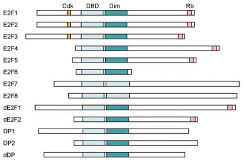
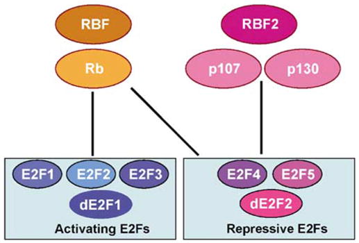
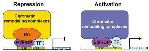
The retinoblastoma gene Rb was the first tumor suppressor gene cloned, and it is well known as a negative regulator of the cell cycle through its ability to bind the transcription factor E2F and repress transcription of genes required for S phase. Although over 100 other proteins have been reported to interact with Rb, in most cases these interactions are much less well characterized. Therefore, this review will primarily focus on Rb and E2F interactions. In addition to cell cycle regulation, studies of Rb and E2F proteins in animal models have revealed important roles for these proteins in apoptosis and differentiation. Recent screens of Rb/E2F target genes have identified new targets in all these areas. In addition, the mechanisms determining how different subsets of target genes are regulated under different conditions have only begun to be addressed and offer exciting possibilities for future research.


Figures

Figure 1
Figure 1
The mammalian and Drosophila pocket protein families. The pocket protein family in mammals consists of Rb, p107 and p130, and in Drosophila contains RBF and RBF2. The pocket domain, responsible for most protein–protein interactions, consists of two conserved sequences, A and B. The spacer region between A and B is conserved between p107 and p130 (yellow box), and can bind to cyclin/cdk complexes. The activity of Rb proteins is controlled by phosphorylation at numerous phosphorylation sites (indicated for Rb and RBF by *).
Figure 2
Figure 2
The mammalian and Drosophila E2F protein families. In mammals the E2F family is composed of E2Fs 1–8, DP1 and DP2, while in Drosophila it consists of dE2F1, dE2F2 and dDP. All E2Fs have a conserved DNA-binding domain (DBD) and (except for E2Fs 7 and 8) a dimerization domain for binding to DP (Dim). The mammalian-activating E2Fs 1–3 contain a cyclin/cdk-binding domain (cdk) at the N-terminus and a strong activation domain at the C-terminus. This activation domain overlaps with the Rb-binding domain (Rb) so that binding by Rb masks the activation domain. The repressive E2Fs 4 and 5 also bind the pocket proteins but lack the cyclin/cdk binding domain. E2F 6 shares only the DBD and dimerization domain with the rest of the family, while E2Fs 7 and 8 have two conserved DBDs, allowing them to function without DP to form a heterodimer. The DP proteins are distantly related members of the family that share the DBD and dimerization domain, allowing them to form heterodimers with E2F proteins.
Figure 3
Figure 3
Interactions between the Rb and E2F proteins in mammals and Drosophila. The mammalian activating E2Fs, E2Fs 1–3, interact primarily with Rb. Similarly, the Drosophilaactivating E2F, dE2F1, interacts only with RBF. The mammalianrepressive E2Fs, E2Fs 4 and 5, interact with p107 and p130. In addition, E2F 4 can also interact with Rb. Similarly, the Drosophila-repressive E2F, dE2F2, interacts with both RBF and RBF2.
Figure 4
Figure 4
Combinatorial control of gene expression by Rb/E2F with other transcription factors. Owing to specific interactions with other transcription factors, individual members of the Rb and E2F families can differentially regulate specific target genes. As shown in the repression model, specific Rb/E2F complexes may be preferentially recruited to an E2F-binding site based on the ability of Rb and/or E2F to bind to another transcription factor with an adjacent binding site. Similarly, in the activation model, the ability of individual E2F family members to bind to other transcription factors may preferentially recruit both activators to promoters containing adjacent binding sites, synergistically activating transcription.

Similar articles

Cited by

References

    1. Apostolova MD, Ivanova IA, Dagnino C, D’Souza SJ, Dagnino L. J Biol Chem. 2002;277:34471–34479. - PubMed
    1. Asano M, Nevins JR, Wharton RP. Genes Dev. 1996;10:1422–1432. - PubMed
    1. Attwooll C, Denchi EL, Helin K. EMBO J. 2004a;23:4709–4716. - PMC - PubMed
    1. Attwooll C, Oddi S, Cartwright P, Prosperini E, Agger K, Steensgaard P, et al. J Biol Chem. 2004b;280:1199–1208. - PubMed
    1. Bosco G, Du W, Orr-Weaver TL. Nat Cell Biol. 2001;3:289–295. - PubMed

Publication types