

Dynamical changing patterns of histological structure and ultrastructure of liver graft undergoing warm ischemia injury from non-heart-beating donor in rats
- PMID: 16937478
- PMCID: PMC4087630
- DOI: 10.3748/wjg.v12.i30.4902
Dynamical changing patterns of histological structure and ultrastructure of liver graft undergoing warm ischemia injury from non-heart-beating donor in rats
Abstract
Aim: To investigate the histological and ultra-structural characteristics of liver graft during different of warm ischemia time (WIT) in rats and to predict the maximum limitation of liver graft to warm ischemia.
Methods: The rats were randomized into 7 groups undergoing warm ischemia injury for 0, 10, 15, 20, 30, 45 and 60 min, respectively. All specimens having undergone warm ischemia injury were investigated dynamically by light and electron microscopy, and histochemistry staining. After orthotopic liver transplantation (OLT), the recovery of morphology of liver grafts after 6, 24 and 48 h was observed.
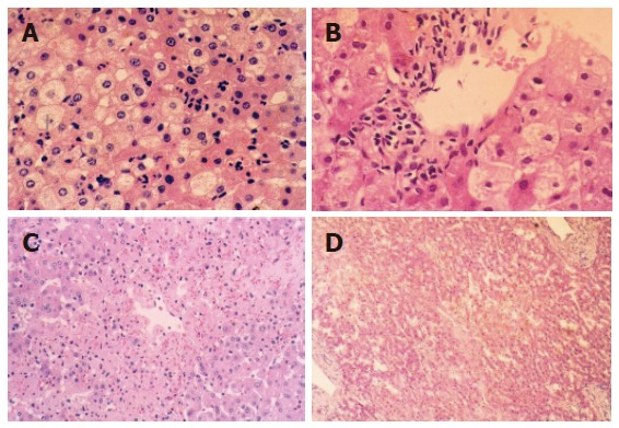
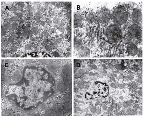
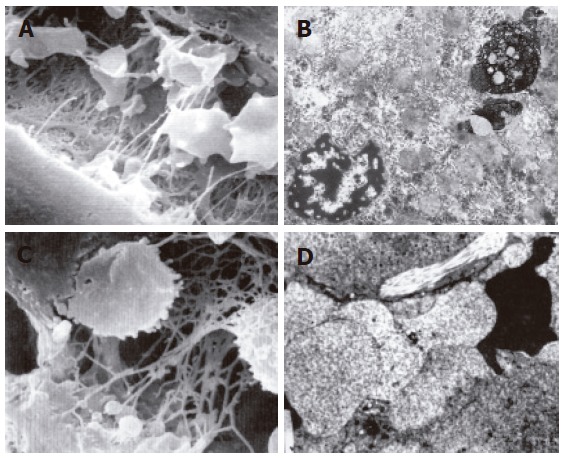
Results: The donor liver from non-heart-beating donors (NHBD) underwent ischemia injury both in the warm ischemia period and in the reperfusion period. Morphological changes were positively related to warm ischemia injury in a time-dependent manner during the reperfusion period. The results demonstrated that different degrees of histiocyte degeneration were observed when WIT was within 30 min, and became more severe with the prolongation of WIT, no obvious hepatocyte necrosis was noted in any specimen. In the group undergoing warm ischemia injury for 45 min, small focal necrosis occurred in the central area of hepatic lobule first. In the group undergoing warm ischemia injury for 60 min, patchy or diffused necrosis was observed and the area was gradually extended, while hepatic sinusoid endothelial cells were obviously swollen. Hepatic sinusoid was obstructed and microcirculation was in disorder.
Conclusion: The rat liver graft undergoing warm ischemia injury is in the reversible stage when the WIT is within 30 min. The 45 min WIT may be a critical point of rat liver graft to endure warm ischemia injury. When the WIT is over 60 min, the damage is irreversible.
Figures
Similar articles
-
Dynamic microcirculatory changes in liver graft from non-heart-beating donor with warm ischemia injury in rat.Hepatobiliary Pancreat Dis Int. 2004 May;3(2):179-82. Hepatobiliary Pancreat Dis Int. 2004. PMID: 15138105
-
Safe time to warm ischemia and posttransplant survival of liver graft from non-heart-beating donors.World J Gastroenterol. 2004 Nov 1;10(21):3157-60. doi: 10.3748/wjg.v10.i21.3157. World J Gastroenterol. 2004. PMID: 15457563 Free PMC article.
-
Energy metabolism and survival of liver grafts from non-heart-beating donor rats with warm ischemia injury.Hepatobiliary Pancreat Dis Int. 2006 Nov;5(4):521-5. Hepatobiliary Pancreat Dis Int. 2006. PMID: 17085336
-
Non beating heart donors as a possible source for liver transplantation.Acta Chir Belg. 2000 Nov-Dec;100(6):268-71. Acta Chir Belg. 2000. PMID: 11236181 Review.
-
An evaluation of the tolerance of the autotransplanted canine lung against warm ischemia.Jpn J Surg. 1989 May;19(3):326-33. doi: 10.1007/BF02471409. Jpn J Surg. 1989. PMID: 2674503 Review.
Cited by
-
The Role of GLP1 in Rat Steatotic and Non-Steatotic Liver Transplantation from Cardiocirculatory Death Donors.Cells. 2019 Dec 9;8(12):1599. doi: 10.3390/cells8121599. Cells. 2019. PMID: 31835410 Free PMC article.
-
The Effect of Fibroblast Growth Factor 15 Signaling in Non-Steatotic and Steatotic Liver Transplantation from Cardiocirculatory Death.Cells. 2019 Dec 14;8(12):1640. doi: 10.3390/cells8121640. Cells. 2019. PMID: 31847428 Free PMC article.
-
Shortening the recipient warm ischemia time could be a strategy for expanding the liver donor pool.World J Gastroenterol. 2025 Mar 7;31(9):103188. doi: 10.3748/wjg.v31.i9.103188. World J Gastroenterol. 2025. PMID: 40061597 Free PMC article.
-
Near-infrared fluorescence imaging with indocyanine green for assessment of donor livers in a rat model of ischemia-reperfusion.BMC Gastroenterol. 2022 Jan 20;22(1):27. doi: 10.1186/s12876-022-02103-5. BMC Gastroenterol. 2022. PMID: 35057742 Free PMC article.
-
Current status and recent advances of liver transplantation from donation after cardiac death.World J Gastrointest Surg. 2011 Nov 27;3(11):167-76. doi: 10.4240/wjgs.v3.i11.167. World J Gastrointest Surg. 2011. PMID: 22180833 Free PMC article.
References
-
- Fondevila C, Busuttil RW, Kupiec-Weglinski JW. Hepatic ischemia/reperfusion injury--a fresh look. Exp Mol Pathol. 2003;74:86–93. - PubMed
-
- Hines IN, Harada H, Wolf R, Grisham MB. Superoxide and post-ischemic liver injury: potential therapeutic target for liver transplantation. Curr Med Chem. 2003;10:2661–2667. - PubMed
-
- Selzner N, Rudiger H, Graf R, Clavien PA. Protective strategies against ischemic injury of the liver. Gastroenterology. 2003;125:917–936. - PubMed
-
- Nuñez JR, Del Rio F, Lopez E, Moreno MA, Soria A, Parra D. Non-heart-beating donors: an excellent choice to increase the donor pool. Transplant Proc. 2005;37:3651–3654. - PubMed
Publication types
MeSH terms
LinkOut - more resources
Full Text Sources
Medical
