

Case Reports
doi: 10.3748/wjg.v12.i31.5075.
Clinical evaluation of submucosal colonic lipomas: decision making
Affiliations
- PMID: 16937511
- PMCID: PMC4087418
- DOI: 10.3748/wjg.v12.i31.5075
Item in Clipboard
Case Reports
Clinical evaluation of submucosal colonic lipomas: decision making
World J Gastroenterol.
.
Abstract
Even lipomas are the most common mesenchymal benign tumors of the gastrointestinal tract, symptomatic colonic presentation is rare. Herein, we evaluated four patients suffering from various size of colonic lipomas and approached by different therapeutic modalities.
Figures
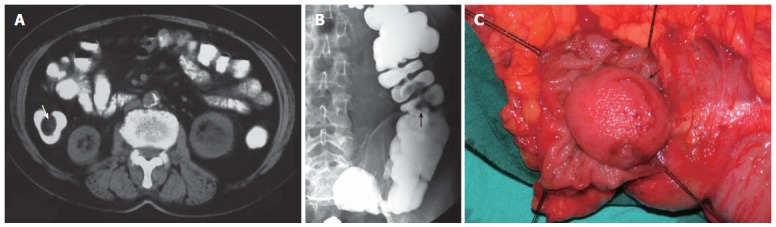
Computed tomography (CT) scan showing a regular contoured 4 cm x 3 cm lesion with fatty density localised in midportion of ascending colon causing a luminal narrowing defect (A), double contrast barium enema showing a filling defect of the protruded polipoid lesion at the hepatic flexura of colon (B), and appearance of the colonic submucosal lipoma during the operation (C).
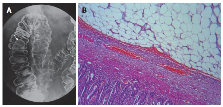
A mass lesion located in the hepatic flexura causing filling defect shown by double contrast enema (A), resected biopsy specimen showing a submucosal benign lipoma composed of mature lipocytes by hematoxylin and eosin staining (B) (x 400).
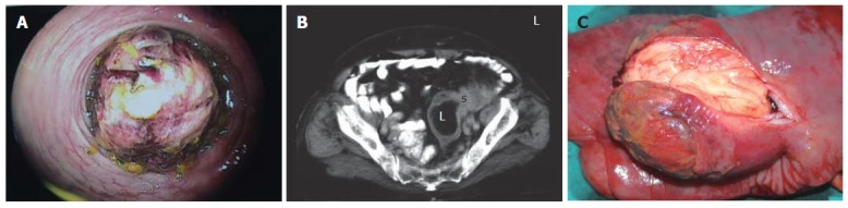
Colonoscopic appearance of a broad- based giant mass acquiring spontaneous hemorrhagic areas and obstructing more than 75% of the lumen of sigmoid colon (A); abdominal computed tomography scans showing the diffuse thickening of sigmoid colon wall, invagination and a distal intraluminal giant mass with fat density with a size of approximately 6 cm x 7 cm (B); macroscopic appearance of the giant sigmoid colon lipoma (C).
Similar articles
-
[Submucosal lipoma of the colon: abdominal cramps with rectal bleeding and weight loss].Ned Tijdschr Geneeskd. 2006 Sep 9;150(36):1990-3. Ned Tijdschr Geneeskd. 2006. PMID: 17002189 Dutch.
-
Giant colonic lipoma presenting with intermittent intestinal obstruction.J Coll Physicians Surg Pak. 2012 Dec;22(12):792-3. J Coll Physicians Surg Pak. 2012. PMID: 23217488
-
Sheep in Wolf's Clothing: Pedunculated Colonic Lipoma with Overlying Hyperplastic and Ulcerated Epithelium.Dig Dis Sci. 2020 Jul;65(7):1951-1953. doi: 10.1007/s10620-020-06188-4. Dig Dis Sci. 2020. PMID: 32157495
-
Giant lipoma of the transverse colon: a case report and review of the literature.Tunis Med. 2009 Jun;87(6):398-402. Tunis Med. 2009. PMID: 19927786 Review.
-
Large colonic lipoma with mucosal ulceration mimicking carcinoma.Gastrointest Endosc. 2003 Sep;58(3):468-70. doi: 10.1067/s0016-5107(03)00035-x. Gastrointest Endosc. 2003. PMID: 14528235 Review. No abstract available.
Cited by
-
Solitary lipoma of ileocaecal valve mimicking Crohn's disease: A case report of a challenging diagnosis for a rare benign tumor of the intestinal tract.Int J Surg Case Rep. 2023 Sep;110:108696. doi: 10.1016/j.ijscr.2023.108696. Epub 2023 Aug 22. Int J Surg Case Rep. 2023. PMID: 37651809 Free PMC article.
-
Ileocecal valve lipoma with refractory hemorrhage.JSLS. 2009 Jan-Mar;13(1):80-3. JSLS. 2009. PMID: 19366548 Free PMC article.
-
Endoscopic submucosal dissection of a large colonic lipoma: Report of two cases.World J Gastroenterol. 2015 Mar 14;21(10):3127-31. doi: 10.3748/wjg.v21.i10.3127. World J Gastroenterol. 2015. PMID: 25780315 Free PMC article.
-
Role of clinical and multidetector computed tomography (MDCT) features in the prediction of patients with intestinal lipoma developing intussusception.Quant Imaging Med Surg. 2024 Jun 1;14(6):3939-3950. doi: 10.21037/qims-23-1530. Epub 2024 May 20. Quant Imaging Med Surg. 2024. PMID: 38846289 Free PMC article.
-
Spontaneous expulsion from rectum: a rare presentation of intestinal lipomas.World J Emerg Surg. 2011 Jun 13;6:19. doi: 10.1186/1749-7922-6-19. World J Emerg Surg. 2011. PMID: 21668995 Free PMC article.
References
-
- Bardají M, Roset F, Camps R, Sant F, Fernández-Layos MJ. Symptomatic colonic lipoma: differential diagnosis of large bowel tumors. Int J Colorectal Dis. 1998;13:1–2. - PubMed
-
- Rogy MA, Mirza D, Berlakovich G, Winkelbauer F, Rauhs R. Submucous large-bowel lipomas--presentation and management. An 18-year study. Eur J Surg. 1991;157:51–55. - PubMed
-
- Vecchio R, Ferrara M, Mosca F, Ignoto A, Latteri F. Lipomas of the large bowel. Eur J Surg. 1996;162:915–919. - PubMed
-
- Bahadursingh AM, Robbins PL, Longo WE. Giant submucosal sigmoid colon lipoma. Am J Surg. 2003;186:81–82. - PubMed
-
- Taylor BA, Wolff BG. Colonic lipomas. Report of two unusual cases and review of the Mayo Clinic experience, 1976-1985. Dis Colon Rectum. 1987;30:888–893. - PubMed
Publication types
MeSH terms
LinkOut - more resources
Full Text Sources
