

Unusual prerectal location of a tailgut cyst: a case report
- PMID: 16937513
- PMCID: PMC4087420
- DOI: 10.3748/wjg.v12.i31.5081
Unusual prerectal location of a tailgut cyst: a case report
Abstract
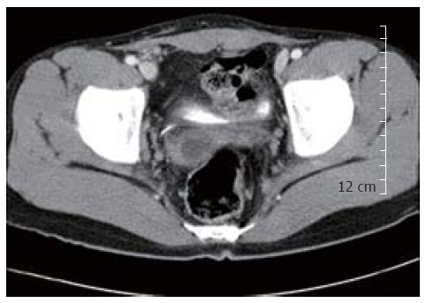
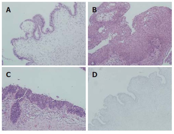
Tailgut cyst is a rare congenital cystic lesion arising from the remnants of the embryonic postanal gut. It occurs exclusively within the retrorectal space and rarely in the perirenal area or in the subcutaneous tissue. A prerectal and retrovesical location of tailgut cyst is extremely rare. To the best of our knowledge, only three cases have been reported in the English literature. We experienced an unusual case of tailgut cyst developed in the prerectal and retrovesical space in a 14-year-old boy. Abdominal computed tomography demonstrated a prerectal cyst which was located at the anterolateral portion to the rectum. The cyst contained yellowish inspissated mucoid material. Microscopically, the cyst was lined by squamous, columnar, cuboidal and transitional epithelia and the wall was fibrotic with dispersed smooth muscle cells. Although tailgut cyst arising in prerectal area is extremely rare, its possibility should be considered in differential diagnosis of a prerectal and retrovesical cystic mass.
Figures
References
-
- Killingsworth C, Gadacz TR. Tailgut cyst (retrorectal cystic hamartoma): report of a case and review of the literature. Am Surg. 2005;71:666–673. - PubMed
-
- Sung MT, Ko SF, Niu CK, Hsieh CS, Huang HY. Perirenal tailgut cyst (cystic hamartoma) J Pediatr Surg. 2003;38:1404–1406. - PubMed
-
- Hjermstad BM, Helwig EB. Tailgut cysts. Report of 53 cases. Am J Clin Pathol. 1988;89:139–147. - PubMed
-
- Mills SE, Walker AN, Stallings RG, Allen MS Jr. Retrorectal cystic hamartoma. Report of three cases, including one with a perirenal component. Arch Pathol Lab Med. 1984;108:737–740. - PubMed
Publication types
MeSH terms
LinkOut - more resources
Full Text Sources
Medical
