

Endoscopic submucosal dissection for stomach neoplasms
- PMID: 16937520
- PMCID: PMC4088006
- DOI: 10.3748/wjg.v12.i32.5108
Endoscopic submucosal dissection for stomach neoplasms
Abstract
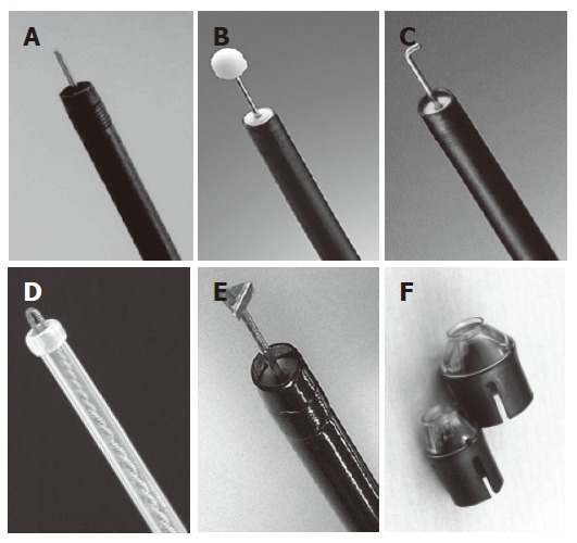
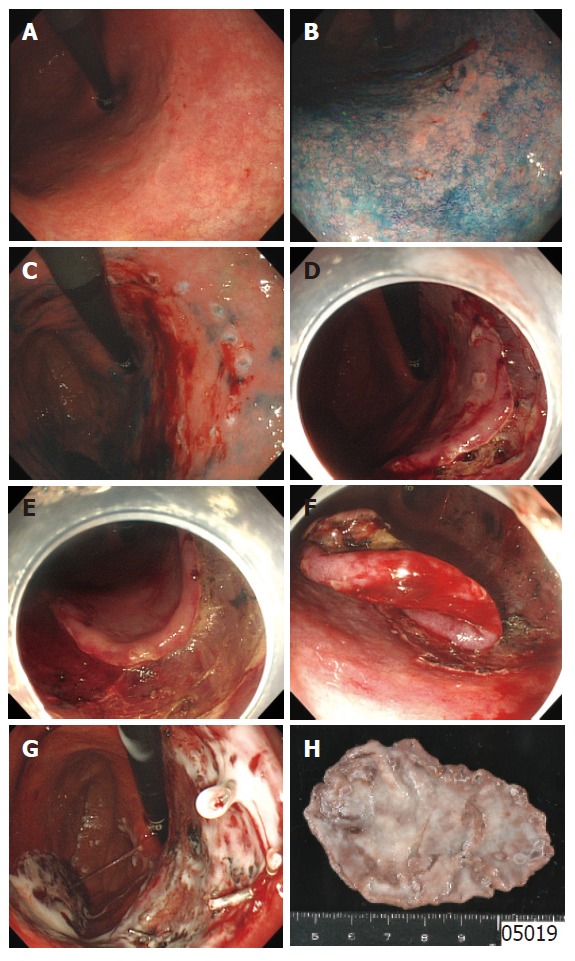
Recent advances in techniques of therapeutic endoscopy for stomach neoplasms are rapidly achieved. One of the major topics in this field is endoscopic submucosal dissection (ESD). ESD is a new endoscopic technique using cutting devices to remove the tumor by the following three steps: injecting fluid into the submucosa to elevate the tumor from the muscle layer, pre-cutting the surrounding mucosa of the tumor, and dissecting the connective tissue of the submucosa beneath the tumor. So the tumors are resectable in an en bloc fashion, regardless of the size, shape, coexisting ulcer, and location. Indication for ESD is strictly confined by two aspects: the possibility of nodal metastases and technical difficulty, which depends on the operators. Although long-term outcome data are still lacking, short-term outcomes of ESD are extremely favourable and laparotomy with gastrectomy is replaced with ESD in some parts of therapeutic strategy for early gastric cancer.
Figures
References
-
- Niwa H. Improvement of fibrogastroscope for biopsy and application of color television and high frequent currents for endoscopic biopsy (in Japanese) Gastroenterol Endosc. 1968;10:31.
-
- Tsuneoka K, Uchida T. Fibergastroscopic polypectomy with snare method and its significance developed in our department - polyp resection and recovery instruments (in Japanese with English abstract) Gastroenterol Endosc. 1969;11:174–184.
-
- Tada M, Shimada M, Murakami F, Shimada M, Mizumachi M, Arima T, Yanai H, Oka S, Shigeeda M, Ogino M, et al. Development of the strip-off biopsy (in Japanese with English abstract) Gastroenterol Endosc. 1984;26:833–839.
-
- Inoue H, Takeshita K, Hori H, Muraoka Y, Yoneshima H, Endo M. Endoscopic mucosal resection with a cap-fitted panendoscope for esophagus, stomach, and colon mucosal lesions. Gastrointest Endosc. 1993;39:58–62. - PubMed
-
- Hirao M, Masuda K, Asanuma T, Naka H, Noda K, Matsuura K, Yamaguchi O, Ueda N. Endoscopic resection of early gastric cancer and other tumors with local injection of hypertonic saline-epinephrine. Gastrointest Endosc. 1988;34:264–269. - PubMed
Publication types
MeSH terms
LinkOut - more resources
Full Text Sources
Medical
Research Materials
Miscellaneous
