Comparative Study
. 2006 Sep 5;103(36):13457-62.
doi: 10.1073/pnas.0604183103. Epub 2006 Aug 24.

Role of T and NK cells and IL7/IL7r interactions during neonatal maturation of lymph nodes

Affiliations
Comparative Study

Role of T and NK cells and IL7/IL7r interactions during neonatal maturation of lymph nodes

Mark C Coles et al. Proc Natl Acad Sci U S A. .

Abstract

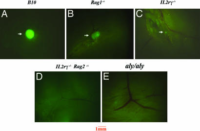
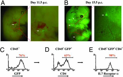
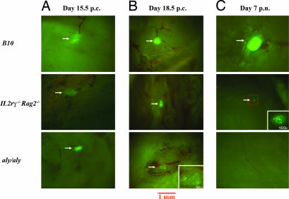
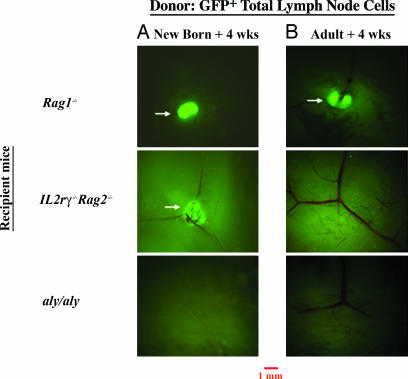
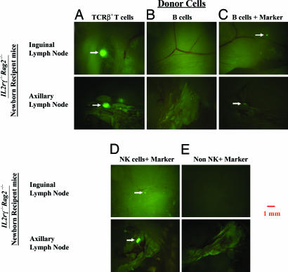
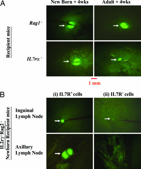
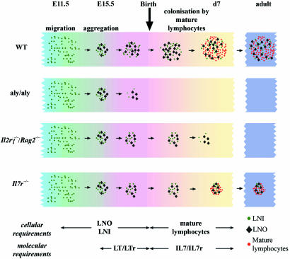
Lymph node (LN) development depends on prenatal interactions occurring between LN inducer and LN organizer cells. We have distinguished defects in LN formation due to failure in embryonic development (aly/aly) from defects in postnatal maturation (Il2rgamma(-/-)Rag2(-/-)). Both mutant strains form normal primordial LNs with differing fate. In aly/aly mice, the LN primordium dissipates irreversibly late in gestation; in contrast, Il2rgamma(-/-)Rag2(-/-) LN anlage persists for a week after birth but disperses subsequently, a process reversible by neonatal transfer of WT IL7r(+) TCR(+) T or natural killer (NK) cells, suggesting a role for IL7/IL7r interactions. Thus, we reveal a unique stage of postnatal LN development during which mature lymphocytes and IL7/IL7r interactions may play an important role.


Conflict of interest statement

Conflict of interest statement: No conflicts declared.

Figures

Fig. 1.
Fig. 1.
Analysis of peripheral LNs in mutant mouse strains crossed to the human CD2 GFP transgenic mice. Inguinal LNs from B10 (A), Rag1−/− (B), Il2rγ−/− (C), Il2rγ−/−Rag2−/− (D), and aly/aly (E) mice transgenic for GFP. Results are representative of 5–10 experiments. Mice were analyzed at 6–8 weeks of age. (Magnification: ×16.)
Fig. 2.
Fig. 2.
Characterization of human CD2 GFP transgenic mice. (A) GFP-expressing cells first appear at sites of LN development (white arrow) at day 11.5 p.c. (B) GFP-expressing cells are found in the developing LN anlagen (cervical, brachial, and inguinal; white arrows) at day 15.5 p.c. (C) Flow cytometric analysis of GFP expression on gated CD45+ cells in developing LN anlagen (day 15.5). (D) CD4 expression on CD45+GFP+ cells. (E) IL7 receptor expression on CD45+GFP+CD4+ cells. Results are representative of 5–10 experiments.
Fig. 3.
Fig. 3.
Analysis of developing LN anlagen from hCD2 GFP, Il2rγ−/−Rag2−/−, and aly/aly mice. (A) Day 15.5 p.c. LN anlage. (B) Day 18.5 p.c. LN anlage. (C) Day 7 p.n. LN. (Magnification: ×32.) (Insets) Images are at magnifications as indicated. Results are representative of 4–10 experiments.
Fig. 4.
Fig. 4.
Rescuing of LN formation by adoptive transfer of lymphocytes in mutant mice. Peripheral LN cells from hCD2-GFP/B6 mice were transferred to into newborn recipient mice (+4 weeks) (A) and adult recipient mice (+4 weeks) (B) of the following genotype: Rag1−/−, Il2rγ−/−Rag2−/−, and aly/aly mice. Analysis of the inguinal LNs is shown 4 weeks after reconstitution. Results are representative of 8–10 experiments. (Magnification: ×16.)
Fig. 5.
Fig. 5.
Rescuing of LN formation by adoptive transfer of specific subsets of lymphocytes in mutant mice. Purified GFP+ T cells (A), YFP+ B cells (B and C), NK cells (D), and non-T/B/NK cells (E) were transferred into newborn IL2rγ−/−Rag2−/− recipient mice. Three weeks later, CFSE-labeled LN cells were transferred i.v. in these mice 72 h before analysis. Images of inguinal and axillary LNs are shown at ×16 magnification. Results in the figure are representative of 3–10 experiments.
Fig. 6.
Fig. 6.
IL7/IL7R interactions are necessary for p.n. rescuing of LNs. (A) Rescuing of LN size in IL7Rα−/− mice by mature WT lymphocytes. WT GFP-expressing LN cells were transferred into Rag1−/− and IL7rα−/− mice. Inguinal LNs are shown 4 weeks after reconstitution from newborn recipients (Left) and adult recipient mice (Right). (B) Rescuing of LN formation by IL7R+ cells from Rag−/− LNs: IL7R+ (Left) and IL7R (Right) cells were transferred into newborn IL2rγ−/−Rag2−/− recipient mice. Three weeks later, CFSE-labeled LN cells were transferred i.v. 72 h before analysis. Images of LNs are shown at ×16 magnification. Results are representative of 10 experiments.
Fig. 7.
Fig. 7.
A model of LN development during the perinatal period. Migrating hemopoietic cells including LNi cells aggregate with LNo cells to form the LN anlage. In normal mice, the structure persists until after birth when it is colonized by mature lymphoid cells generated in the thymus and the bone marrow. In aly/aly mice, these structures degenerate near the end of pregnancy, so that there is no structure to be colonized by mature lymphoid cells after birth. In Il2rγ−/−Rag2−/− mice, these structures persist for a few days after birth, but, because of the complete absence of lymphoid cells, they degenerate within the first week after birth. In Il7r−/− mice, these structures are maintained by the few lymphoid cells that exist in these mice; however, the size of the LNs remains small throughout life.

References

    1. Nishikawa S., Honda K., Vieira P., Yoshida H. Immunol. Rev. 2003;195:72–80. - PubMed
    1. Cupedo T., Kraal G., Mebius R. E. Immunol. Rev. 2002;189:41–50. - PubMed
    1. Cupedo T., Mebius R. E. J. Immunol. 2005;174:21–25. - PubMed
    1. Mebius R. E. Nat. Rev. Immunol. 2003;3:292–303. - PubMed
    1. Wang J. H., Nichogiannopoulou A., Wu L., Sun L., Sharpe A. H., Bigby M., Georgopoulos K. Immunity. 1996;5:537–549. - PubMed

Publication types

LinkOut - more resources