. 2006 Aug 29;103(35):13162-7.
doi: 10.1073/pnas.0605676103. Epub 2006 Aug 22.

Estrogen receptor (ER)-beta isoforms: a key to understanding ER-beta signaling

Affiliations

Estrogen receptor (ER)-beta isoforms: a key to understanding ER-beta signaling

Yuet-Kin Leung et al. Proc Natl Acad Sci U S A. .

Erratum in

  • Proc Natl Acad Sci U S A. 2006 Oct 3;103(40):14977

Abstract

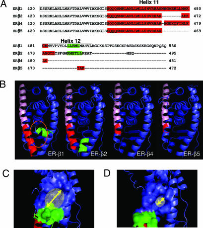
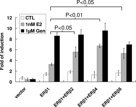
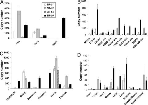
Estrogen receptor beta (ER-beta) regulates diverse physiological functions in the human body. Current studies are confined to ER-beta1, and the functional roles of isoforms 2, 4, and 5 remain unclear. Full-length ER-beta4 and -beta5 isoforms were obtained from a prostate cell line, and they exhibit differential expression in a wide variety of human tissues/cell lines. Through molecular modeling, we established that only ER-beta1 has a full-length helix 11 and a helix 12 that assumes an agonist-directed position. In ER-beta2, the shortened C terminus results in a disoriented helix 12 and marked shrinkage in the coactivator binding cleft. ER-beta4 and -beta5 completely lack helix 12. We further demonstrated that ER-beta1 is the only fully functional isoform, whereas ER-beta2, -beta4, and -beta5 do not form homodimers and have no innate activities of their own. However, the isoforms can heterodimerize with ER-beta1 and enhance its transactivation in a ligand-dependent manner. ER-beta1 tends to form heterodimers with other isoforms under the stimulation of estrogens but not phytoestrogens. Collectively, these data support the premise that (i) ER-beta1 is the obligatory partner of an ER-beta dimer, whereas the other isoforms function as variable dimer partners with enhancer activity, and (ii) a single functional helix 12 in a dimer is sufficient for gene transactivation. Thus, ER-beta behaves like a noncanonical type-I receptor, and its action may depend on differential amounts of ER-beta1 homo- and heterodimers formed upon stimulation by a specific ligand. Our findings have provided previously unrecognized directions for studying ER-beta signaling and design of ER-beta-based therapies.


Conflict of interest statement

Conflict of interest statement: No conflicts declared.

Figures

Fig. 1.
Fig. 1.
Protein sequence analysis and molecular modeling of ER-β isoforms. (A) Protein sequence alignment of the C-terminal regions of ER1, -β2, -β4, and -β5 by using the Clustalw alignment program. The ligand binding domain of ER1 is boxed. The protein sequence forming helix 11 in each isoform is shown in red, whereas the protein sequence participating in helix 12 is in green. (B) Molecular models of ER-β isoforms. The common helix 11 region of each isoform is labeled in pink, whereas the isoform-specific region of helix 11 is highlighted in dark red. The orientation of helix 12 (green) in ER-β2 is different from that of ER-β1, which has “tight” configuration in the ER-β1 binding pocket (orange oval). (C and D) Molecular models of ER-β1 (C) and ER-β2 (D) show the coactivator binding pocket created by electrostatic potential of the amino acid residues in helices 3–5 and 12. The size of the coactivator binding pocket in ER-β2, which is indicated by a yellow arrow, was determined, by using PyMol software, to be smaller than that of ER-β1.
Fig. 2.
Fig. 2.
Characterization of ER-β isoforms. (A) Western blot analysis of ER-β isoforms overexpressed in HEK293 cells and yeast. N-terminal-specific polyclonal ER-β antibody (H150 from Santa Cruz Biotechnology), which would recognize all ER-β isoforms, was used in this study. An equal amount (50 μg) of protein was loaded to each lane. Mock-transfected cells or an untransformed yeast strain were set up as a control experiment (CTL). Samples expressing ER-β1, -β2, -β4, and -β5 were labeled as 1, 2, 4, and 5, respectively. The size of the ER-β isoforms was consistent with the predicted molecular size, ranging from 53 to 59 kDa. Coexpression of ER-β isoforms with ER-β1 was also performed in both cell line and yeast. Lanes 1 and 2, 1 and 4, and 1 and 5 represent the samples overexpressing ER-β1 and -β2, ER-β1 and -β4, and ER-β1 and -β5, respectively. (B) Tabulated results of in vitro estrogen receptor binding assay. Four hundred micrograms of total yeast lysate expressing ER-β isoforms was applied to each binding reaction as described in Materials and Methods. Binding data were calculated and analyzed with GraphPad Prism 4.0 software to determine the Bmax and Kd of each isoform. (C) Effects of SRC-1 on the transactivation activities of ER-β isoforms. SRC-1 expression vector was transfected into HEK293 cells carrying different ER-β isoform expression vectors with the reporter plasmid. Transactivation assays were performed as described in Materials and Methods in the presence or absence of 1 nM E2. Three independent experiments were performed and averaged. The standard deviation was calculated. (D) Dimerization of ER-β isoforms by Y2H experiment. E2 at two different concentrations (1 nM to 1 μM) was incubated overnight with different yeast strains. A Beta-Glo assay was performed to quantify the reporter (β-gal) activity. The higher the reporter activity, the stronger the interaction between two of the same ER-β (homodimer) or different (heterodimer) isoforms. Four types of homodimers (β1 + β1, β2 + β2, β4 + β4, and β5 + β5) and three kinds of heterodimers (β1 + β2, β1 + β4, and β1 + β5) were subjected to Y2H analyses. The background value was subtracted during data analyses. Experiments were performed in triplicate, and the standard deviation was calculated. All results were summarized in this figure, except for β2, β4, and β5 homodimers, in which activities were undetectable.
Fig. 3.
Fig. 3.
Transactivation activities of ER-β homo- and heterodimer in HEK293 cells. Single and double transfection of ER-β isoform expression vectors were performed to study the effects of ER-β homo- and heterodimers, respectively, on the transactivation of ERE reporter. E2, BPA, EE2, DES, genistein, and apigenin at different concentrations (100 pM to 1 μM) were incubated at 24 h after transfection. A control experiment was set up with vehicle. After 24 h of incubation, a Bright-Glo assay was used to measure the luciferase activity. Three independent experiments were performed and averaged. The standard deviation was calculated. A Student t test was applied to determine the significance between ER-β isoform coexpression with ER-β1 and ER-β1 alone with the same treatment. ∗∗, P < 0.01.
Fig. 4.
Fig. 4.
Real-time PCR analysis of pS2 gene expression. HEK293 cells were transfected with ER1, a combination of ER1 with ER2/4/5, or an empty vector as a control. After 24 h of transfection, the cells were treated with 1 nM E2 or 1 μM genistein for another 24 h. The change in pS2 expression level was monitored by real-time PCR analysis. Three independent experiments were performed and averaged. The standard deviation was calculated. A Student t test was applied to determine the significance between ER-β isoforms coexpression with ER-β1 and ER-β1 alone with the same treatment.
Fig. 5.
Fig. 5.
Tissue distribution of ER-β isoforms in various human cell lines (A and B) and human tissues (C and D) by real-time PCR analyses. Transcript levels of each isoform were determined by real-time PCR and expressed as “number of copy” (see Materials and Methods). The expression level of ER-β isoforms was normalized by human GAPDH gene level. Three independent experiments were performed and averaged. The standard deviations were calculated and shown as error bars.
Fig. 6.
Fig. 6.
Model for ER-β isoforms interaction during transcription. (Upper) Putative protein recruitment by an ER-β1 homodimer and heterodimer. Comediators/cointegrators (M1–5) or coactivators (CoAct) with various numbers of nuclear receptor boxes (NR) recruited by ER-β homo- and heterodimers may be different. (Lower) The putative ultimate transcriptional machinery complex recruited by an ER-β heterodimer in a hypothetical promoter. A set of coregulators (M1–5), which maintains an active form of nucleosomes through acetylation (“Ac”-labeled purple spheres) and functions to form an active transcriptional complex with RNA polymerase II (RNA pol), is congregated in an asymmetrical manner around the ER-β1-βn heterodimer, which binds to a symmetrical palindromic ERE.

References

    1. Kuiper G. G., Enmark E., Pelto-Huikko M., Nilsson S., Gustafsson J. A. Proc. Natl. Acad. Sci. USA. 1996;93:5925–5930. - PMC - PubMed
    1. Koehler K. F., Helguero L. A., Haldosen L. A., Warner M., Gustafsson J. A. Endocr. Rev. 2005;26:465–478. - PubMed
    1. Zhu X., Leav I., Leung Y. K., Wu M., Liu Q., Gao Y., McNeal J. E., Ho S. M. Am. J. Pathol. 2004;164:2003–2012. - PMC - PubMed
    1. Evans R. M. Science. 1988;240:889–895. - PMC - PubMed
    1. Kraichely D. M., Sun J., Katzenellenbogen J. A., Katzenellenbogen B. S. Endocrinology. 2000;141:3534–3545. - PubMed

Publication types

MeSH terms

Associated data

LinkOut - more resources