. 2006 Sep 5;103(36):13391-6.
doi: 10.1073/pnas.0605950103. Epub 2006 Aug 25.

The Nck-interacting kinase phosphorylates ERM proteins for formation of lamellipodium by growth factors

Affiliations

The Nck-interacting kinase phosphorylates ERM proteins for formation of lamellipodium by growth factors

Martin Baumgartner et al. Proc Natl Acad Sci U S A. .

Abstract

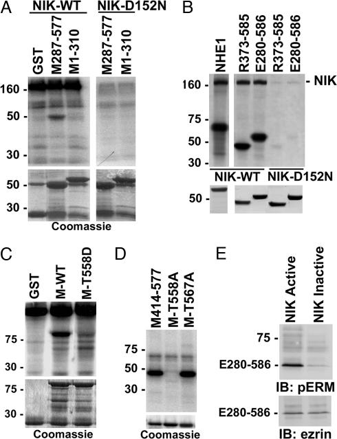
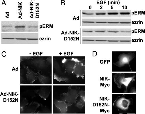
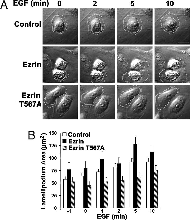
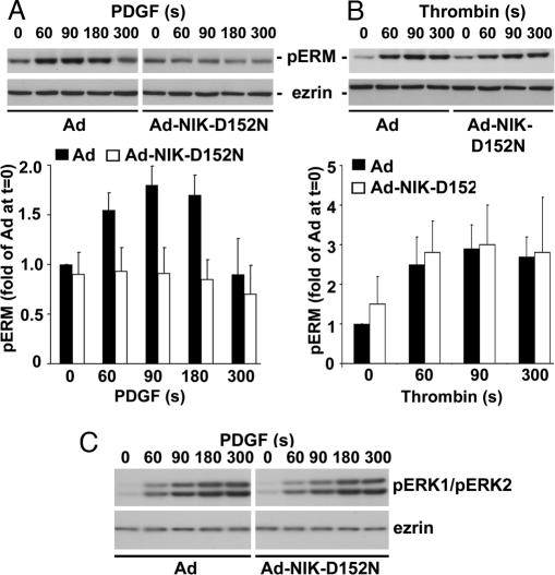
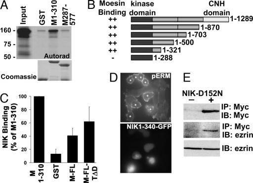
The mammalian Ste20-like Nck-interacting kinase (NIK) and its orthologs Misshapen in Drosophila and Mig-15 in Caenorhabditis elegans have a conserved function in regulating cell morphology, although through poorly understood mechanisms. We report two previously unrecognized actions of NIK: regulation of lamellipodium formation by growth factors and phosphorylation of the ERM proteins ezrin, radixin, and moesin. ERM proteins regulate cell morphology and plasma membrane dynamics by reversibly anchoring actin filaments to integral plasma membrane proteins. In vitro assays show that NIK interacts directly with ERM proteins, binding their N termini and phosphorylating a conserved C-terminal threonine. In cells, NIK and phosphorylated ERM proteins localize at the distal margins of lamellipodia, and NIK activity is necessary for phosphorylation of ERM proteins induced by EGF and PDGF, but not by thrombin. Lamellipodium extension in response to growth factors is inhibited in cells expressing a kinase-inactive NIK, suppressed for NIK expression with siRNA oligonucleotides, or expressing ezrin T567A that cannot be phosphorylated. These data suggest that direct phosphorylation of ERM proteins by NIK constitutes a signaling mechanism controlling growth factor-induced membrane protrusion and cell morphology.


Conflict of interest statement

Conflict of interest statement: No conflicts declared.

Figures

Fig. 1.
Fig. 1.
NIK activity is necessary for extension of lamellipodium by EGF. (A) Time-lapse images of MTLn3 cells infected with empty Ad or Ad expressing WT NIK (Ad-NIK) or NIK-D152N (Ad-NIK-D152N) and treated with EGF (25 nM) are shown for the indicated times. See also Movies 1–3. White lines drawn around the periphery of cell bodies and the distal margins of membrane protrusions were used to calculate lamellipodium area. (B) Means ± SEM of lamellipodia area obtained from six cells in three cell preparations are shown. (C) Representative kymographs from cells infected with the indicated Ad and treated with EGF are shown. Arrows indicate maximum membrane protrusion. (Scale bar: 10 μm.)
Fig. 2.
Fig. 2.
NIK directly phosphorylates the C terminus of ERM proteins. (A) Autoradiograms of kinase reaction (Upper) and Coomassie-stained fusion proteins (Lower) are shown. Myc-tagged WT NIK or NIK-D152N was expressed in COS-7 cells, immunoprecipitated, and used for in vitro kinase assays with GST or GST-fusion proteins containing moesin C terminus (M287-577) or N terminus (M1-310). The high-molecular-mass band at 160 kDa corresponds to autophosphorylated NIK. (B) Autoradiograms of kinase reaction (Upper) and Coomassie-stained fusion proteins (Lower) are shown. WT NIK or NIK-D152N was expressed, immunoprecipitated, and reacted in vitro with GST fusion proteins containing the C terminus of NHE1, radixin (R373-585), or ezrin (E280-586). (C) Autoradiograms of kinase reaction with WT NIK as in A and B (Upper) and Coomassie-stained fusion proteins (Lower) are shown. GST and fusion proteins containing full-length moesin WT (M-WT) or moesin with T558 substituted with aspartic acid (M-T558D) were used as substrates. (D Upper) Autoradiogram of kinase reaction with purified NIK domain (amino acids 22–374) and fusion proteins of the C terminus of moesin WT (M414–577) or containing single substitutions of Thr-558 or Thr-567 with alanine (M-T558A and M-T567A, respectively) is shown. (D Lower) Coomassie-stained fusion proteins used for kinase reaction are shown. (E) Immunoblots (IB) with antiphospho-ERM antibodies (Upper) and antiezrin antibodies (Lower) of C-terminal ezrin translated in vitro and incubated with purified NIK domain active or inactivated by boiling are shown.
Fig. 3.
Fig. 3.
NIK activity is necessary for phosphorylation of ERM proteins by EGF. (A) Immunoblots for pERM proteins (Upper) and ezrin (Lower) in lysates of quiescent MTLn3 cells infected with Ad, Ad-NIK, or Ad-NIK-D152N are shown. (B) Time-dependent phosphorylation of ERM proteins with EGF is indicated by immunoblotting for pERM proteins and ezrin in lysates of MTLn3 cells infected with Ad or Ad-NIK-D152N. (C) Immunolabeling with anti-pERM antibodies of MTLn3 cells infected with Ad or Ad-NIK-D152N in the absence (−EGF) or presence of 25 nM EGF for 5 min (+EGF) indicates pERM proteins are predominantly in lamellipodia in Ad-infected cells with EGF. (D) GFP fluorescence (Top) and anti-Myc immunolabeling of cells expressing NIK-Myc (Middle) or NIK-D152N-Myc (Bottom) are shown. (Scale bar: 10 μm.)
Fig. 4.
Fig. 4.
Phosphorylation of T567 in ezrin is necessary for lamellipodium extension with EGF. (A) Time-lapse images of MTLn3 cells transfected with empty vector (Control) or vector encoding WT ezrin (Ezrin) or ezrin T567A (EzrinT567A) and treated with EGF (25 nM) are shown for the indicated times. See also Movies 4–6. Transfected cells (outlined in white) were identified by coexpression of GFP. Lamellipodium area was determined as in Fig. 1. (B) Mean ± SEM of lamellipodium area of six cells in three cell preparations is shown. (Scale bar: 10 μm.)
Fig. 5.
Fig. 5.
NIK activity is necessary for phosphorylation of ERM proteins by PDGF but not by thrombin. Representative immunoblots of lysates prepared from CCL39 fibroblasts infected with Ad or Ad-NIK-D152N and treated for the indicated times with PDGF (50 ng/ml) (A Upper and C) or thrombin (30 nM) (B Upper) are shown. Blots were probed with anti-pERM antibodies (A and B), antiphospho-ERK antibodies (C), or antiezrin antibodies (AC). Data normalized from five (A Lower) and three (B Lower) cell preparations are expressed as the mean ± SEM of fold-change relative to abundance of pERM proteins in Ad-infected cells at time 0. The immunoblot in C is representative of five cell preparations.
Fig. 6.
Fig. 6.
NIK 1-310 binds the FERM domain of moesin and is sufficient to phosphorylate ERM proteins in cells. (A Upper) Autoradiogram of binding assay with full-length NIK translated in vitro in the presence of [35S]methionine and GST and GST-moesin FERM domain (M1-310) and C terminus (M287-577) is shown. (A Lower) Coomassie-stained fusion proteins are shown. (B) NIK binding to the indicated fusion proteins relative to binding GST-M1-310 is shown. (C) Schematic representation of full-length (resides 1–1289) and C-terminal-truncated NIK used in binding assays is shown. ++, domains with comparable binding to GST-moesin 1–310. (D) Expression of an EGFP fusion of truncated NIK-1-340 is sufficient to increase phosphorylation of endogenous ERM proteins in CCL39 fibroblasts. Phosphorylation of ERM proteins was determined by immunolabeling with anti-pERM antibody. *, cells expressing NIK-1-340-EGFP are identified by fluorescence. (E) Ezrin coprecipitates with Myc-tagged kinase-inactive NIK (mNIK-D152N). (Top and Middle) Myc immunoprecipitates from lysates of CCL39 fibroblasts transfected with empty vector (−) or vector containing Myc-NIK-D152N (+) were probed with antibodies against Myc (Top) and ezrin (Middle). (Bottom) Ezrin in cell lysates was determined by immunoblotting. IP, immunoprecipitation; IB, immunoblotting.

References

    1. Su Y. C., Han J., Xu S., Cobb M., Skolnik E. Y. EMBO J. 1997;16:1279–1290. - PMC - PubMed
    1. Fu C. A., Shen M., Huang B. C., Lasaga J., Payan D. G., Luo Y. J. Biol. Chem. 1999;274:30729–30737. - PubMed
    1. Hu Y., Leo C., Yu S., Huang B. C., Wang H., Shen M., Luo Y., Daniel-Issakani S., Payan D. G., Xu X. J. Biol. Chem. 2004;279:54387–54397. - PubMed
    1. Nakano K., Kanai-Azuma M., Kanai Y., Moriyama K., Yazaki K., Hayashi Y., Kitamura N. Exp. Cell Res. 2003;287:219–227. - PubMed
    1. Su Y. C., Maurel-Zaffran C., Treisman J. E., Skolnik E. Y. Mol. Cell Biol. 2000;20:4736–4744. - PMC - PubMed

Publication types

MeSH terms