Review
. 2006 Sep 29;361(1473):1463-75.
doi: 10.1098/rstb.2006.1886.

Cell replacement therapy in neurological disease

Affiliations
Review

Cell replacement therapy in neurological disease

Steven A Goldman et al. Philos Trans R Soc Lond B Biol Sci. .

Abstract

Diseases of the brain and spinal cord represent especially daunting challenges for cell-based strategies of repair, given the multiplicity of cell types within the adult central nervous system, and the precision with which they must interact in both space and time. Nonetheless, a number of diseases are especially appropriate for cell-based therapy, in particular those in which single phenotypes are lost, and in which the re-establishment of vectorially specific connections is not entirely requisite for therapeutic benefit. We review here a set of potential therapeutic indications that meet these criteria as potentially benefiting from the transplantation of neural stem and progenitor cells. These include: (i) transplantation of phenotypically restricted neuronal progenitor cells into diseases of a single neuronal phenotype, such as Parkinson's disease; (ii) implantation of mixed progenitor pools into diseases characterized by the loss of a limited number of discrete phenotypes, such as spinal cord injury and the motor neuronopathies; (iii) transplantation of glial and nominally oligodendrocytic progenitor cells as a means of treating disorders of myelin; and (iv) transplantation of neural stem cells as a means of treating lysosomal storage disorders and other diseases of enzymatic deficiency. Among the diseases potentially approachable by these strategies, the myelin disorders, including the paediatric leucodystrophies as well as adult traumatic and inflammatory demyelinations, may present the most compelling targets for cell-based neurological therapy.


Figures
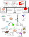
Figure 1
Figure 1
Sources, isolation and use of defined progenitor phenotypes. This figure schematizes the methods of isolating prospectively defined neural progenitor phenotypes from a variety of human cell and tissue sources, and highlights several of the experimental purposes to which these cells may be allocated and devoted. EGFP, enhanced green fluorescent protein; MAGS, magnetic activated cell sorting; PSA-NCAM; polysialylated neural cell adhesion molecule; LTR, long terminal repeat. Adapted from Goldman (2005b).
Figure 2
Figure 2
Myelination by engrafted OPCs. (a–c) Implanted human foetal OPCs myelinated extensive regions of shiverer mouse forebrain. (a, b) MBP expression by sorted human OPCs, implanted into neonatal shiverer mice, indicates that large regions of the corpus callosum ((a) and (b), different mice) have myelinated by 12 weeks (MBP, green). (c) Donor-derived myelin extended throughout the internal capsules. (d–h) New myelin was exclusively derived from human donor cells. (d) MBP (green), in a shiverer callosum three months after neonatal graft, is associated with human donor cells, identified by human nuclear antigen (hNA, red). (e–h) Confocal images of implanted shiverer callosum, with human cells (hNA, red) surrounded by associated MBP (green). ((e) Merged images of (f–h), 1 μm apart.) (i) OPCs were recruited as oligodendrocytes or astrocytes in a context-dependent manner, such that they matured as MBP+ oligodendrocytes in the white matter, but as glial fibrillary acidic protein (GFAP+) astrocytes in both white and grey matter. Panel (i) shows the striatocallosal border of a shiverer brain, three months after human foetal OPC transplant (hNA, blue). Donor-derived MBP (red) fills the callosum, while donor-derived GFAP+ (green) astrocytes predominate in the striatum and ventricular wall. Scale bar, 1 mm (a–c), 100 μm (d), 20 μm (e–h), 200 μm (i). Adapted from Goldman (2005b) and Windrem et al. (2004).
Figure 3
Figure 3
Dual site cell transplantation. Widespread infiltration of human donor OPCs into the shiverer brain and brainstem follows neonatal injection into the cisterna magnum and corpus callosum. (a) This animal was injected on P0 with 1×10⁵ cells each into the cisterna magnum(*), and corpus callosum (CC), then sacrificed at day 30 and stained for human nuclear antigen (red, arrow) to identify donor cells. SC, superior colliculus. (b) This animal was treated identically as that in (a), except that it was given an additional injection into the cerebellar central white matter. It was sacrificed two months later, by which time it exhibited not only widespread forebrain and brainstem cell dispersal, but also early MBP production throughout the cerebellar white matter (green). (c) An assessment of five different measures of engraftment success, derived from another set of shiverers sacrificed at 12 weeks, and assessed by selectively scoring areas of robust myelination, so as to estimate maximal efficacy of engraftment. Scored metrics include: (c-1) proportion of human nuclear antigen (hN+) donor cells expressing MBP; (c-2) density of myelinating donor cells per mm3; (c-3) number of axons ensheathed per donor cell and per MBP+ donor cell; (c-4) donor cell density at 12 weeks, compared between fimbria, callosum and cerebellar white matter (WM).
Figure 4
Figure 4
Axonal ensheathment and myelin compaction by engrafted human progenitor cells. (a) A confocal micrograph showing a triple immunostain for MBP (red), human nuclear antigen (blue) and neurofilament protein (green). In this image, all MBP immunostaining is derived from the sorted human OPCs, whereas the NF+ axons are those of the mouse host. Arrows identify murine axons ensheathed by human MBP. (b) A 2 μm deep composite of optical sections, taken through the corpus callosum of a shiverer recipient sacrificed 12 weeks after foetal OPC implantation. Shiverer axons were scored as ensheathed when the yellow index lines intersected an NF+ axon abutted on each side by MBP. The asterisk indicates the field enlarged in the inset. (c, d) Representative electron micrographs of 16-week-old shiverer homozygotes, implanted with human OPCs shortly after birth. The images show shiverer axons ensheathed by densely compacted myelin. The asterisk indicates the field enlarged in the inset. Inset: major dense lines are noted between lamellae, providing electron microscopic confirmation of myelination. (e) High power confocal images of MBP+ donor-derived myelin sheaths (green) at internodal junctions, characterized by expression of Caspr protein (red) at the paranodal borders. Left, a single 0.4 μm optical section; right, a z-stack composite. Caspr staining confirmed nodes of Ranvier between adjacent donor-derived myelinated segments (Einheber et al. 1997); these results suggest physiologically appropriate conduction support by donor-derived myelin. Scale bar, 20 μm (a), 40 μm (b), 1 μm (c, d). Adapted from Goldman (2005b) and Windrem et al. (2004).
Figure 5
Figure 5
Foetal and adult OPCs differed substantially in their speed and efficiency of myelinogenesis. (a) Adult-derived human OPCs (hNA, red) achieved dense MBP (green) by four weeks after graft. (b) In contrast, foetal OPCs expressed no MBP at four weeks, and none until 12 weeks. (c, d) Low- and high-power images of the callosal–fimbrial junction of a shiverer brain; dense myelination by 12 weeks after perinatal delivery of adult OPCs. (e, f) Adult OPCs developed mature myelin ultrastructure and major dense lines within five weeks of perinatal injection. (e, f) Myelin in a shiverer homozygote five weeks after perinatal injection of adult OPCs. Mice injected with foetal OPCs exhibited no evidence of myelination at this time point. (g) The distribution of hNA+ adult OPCs (red), four weeks after implant. (h) A higher proportion of adult OPCs developed MBP expression than did foetal OPCs, when assessed 12 weeks after transplant. (i) Foetal OPCs nonetheless engrafted more efficiently and in higher numbers than did adult OPCs. *p<0.05; **p<0.005, Student's t-test. (j) A plot comparing the number of ensheathed axons per donor cell achieved by foetal and adult-derived OPCs. Ensheathment was defined by confocal-imaged MBP+ enwrapping NF+ axons, and was measured as a function of total donor cell number (left), and of MBP+ donor-derived oligodendrocytes (right). The difference between foetal and adult donor ensheathment efficiencies was significant by Mann–Whitney (p<0.02). Scale bar, 100 μm (a, b), 1 mm (c), 30 μm (d), 1 μm (e, f). Adapted from Windrem et al. (2004).

Similar articles

Cited by

References

    1. Arber S, Han B, Mendelsohn M, Smith M, Jessell T.M, Sockanathan S. Requirement for the homeobox gene Hb9 in the consolidation of motor neuron identity. Neuron. 1999;23:659–674. 10.1016/S0896-6273(01)80026-X - DOI - PubMed
    1. Back S, Rivkees S. Emerging concepts in periventricular white matter injury. Semin. Perinatol. 2004;6:405–414. 10.1053/j.semperi.2004.10.010 - DOI - PubMed
    1. Bjorklund A, Lindvall O. Cell replacement therapies for central nervous system disorders. Nat. Neurosci. 2000;3:537–544. 10.1038/75705 - DOI - PubMed
    1. Bjorklund L, Sanchez-Pernaute R, Chung S, Jenkins B, Wahlestedt C, Kim K.M, Isacson O. Embryonic stem cells develop into functional dopaminergic neurons after transplantation in a Parkinson rat model. Proc. Natl Acad. Sci. USA. 2002;99:2344–2349. 10.1073/pnas.022438099 - DOI - PMC - PubMed
    1. Brustle O, Choudhary K, Karram K, Huttner A, Murray K, Dubois-Dalcq M, McKay R. Chimeric brains generated by intraventricular transplantation of fetal human brain cells into embryonic rats. Nat. Biotechnol. 1998;16:1040–1044. - PubMed

Publication types

MeSH terms

LinkOut - more resources