Review
. 2006 Sep 29;361(1473):1575-92.
doi: 10.1098/rstb.2006.1882.

Intracellular control of developmental and regenerative axon growth

Affiliations
Review

Intracellular control of developmental and regenerative axon growth

Feng-Quan Zhou et al. Philos Trans R Soc Lond B Biol Sci. .

Abstract

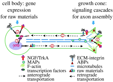
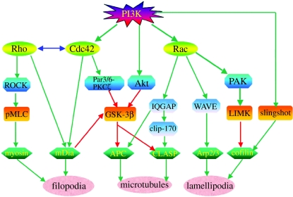
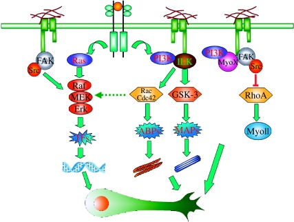
Axon growth is a highly regulated process that requires stimulating signals from extracellular factors. The extracellular signals are then transduced to regulate coordinately gene expression and local axon assembly. Growth factors, especially neurotrophins that act via receptor tyrosine kinases, have been heavily studied as extracellular factors that stimulate axon growth. Downstream of receptor tyrosine kinases, recent studies have suggested that phosphatidylinositol-3 kinase (PI3K) regulates local assembly of axonal cytoskeleton, especially microtubules, via glycogen synthase kinase 3beta (GSK-3beta) and multiple microtubule binding proteins. The role of extracellular signal regulated kinase (ERK) signalling in regulation of local axon assembly is less clear, but may involve the regulation of local protein translation. Gene expression during axon growth is regulated by transcription factors, among which cyclic AMP response element binding protein and nuclear factors of activated T-cells (NFATs) are known to be required for neurotrophin (NT)-induced axon extension. In addition to growth factors, extracellular matrix molecules and neuronal activity contribute importantly to control axon growth. Increasingly, evidence suggests that these influences act to enhance growth via coordinating with growth factor signalling. Finally, evidence is emerging that developmental versus regenerative axon growth may be mediated by distinct signalling pathways, both at the level of gene transcription and at the level of local axon assembly.


Figures

Figure 1
Figure 1
A coordinated genetic programme and local signalling cascade regulate axon growth. To mediate efficient axon assembly, extracellular axon growth promoting factors, such as neurotrophins and ECMs, (1) activate signalling cascades locally at the axon that eventually converge to regulate actions of actin binding proteins (ABPs) and microtubule associated proteins (MAPs). These cytoskeletal-associated molecules then mediate axon assembly via orchestrated modulation of actin and microtubule polymerization. At the same time, (2) activated signalling mediators can be retrogradely transported from the axon to the cell body. After arriving at the soma, these signal mediators initiate (3) activation of a set of transcription factors that control axon growth. Extracellular factors may also activate transcription factors directly at the soma to induce gene expression. Finally, gene expression mediated by these transcription factors and subsequent protein translation produce the raw materials for new axons, including cytoskeletal elements. (4) These raw materials are then transported anterogradely and incorporated into the growing axon.
Figure 2
Figure 2
Regulation of the axon cytoskeleton by PI3K signalling. Small GTPases, including Rac, Cdc42 and Rho, are major downstream mediators of PI3K signalling that regulate the cytoskeleton. Both Rac and Cdc42 are activated by neurotrophin signalling downstream of PI3K. Activated Rac can activate WAVE proteins and subsequently the Arp2/3 complex to promote actin polymerization that mediates lamellipodia formation. Rac can also activate PAK and LIM kinase (LIMK), which phosphorylates and inactivates the actin depolymerization factor cofilin. Cofilin is activated by dephosphorylation via the phosphatase Slingshot directly downstream of PI3K. Appropriate regulation of cofilin activity is important for actin polymerization. Cdc42 is mainly involved in regulation of actin filopodial formation, probably via the actin binding protein mDia. In addition to regulation of actin filaments, both Rac and Cdc42 regulate microtubule assembly and dynamics. Activated Rac at the lamellipodial leading edge can capture and stabilize dynamic microtubules via the interaction of its effector IQGAP1 with the microtubule plus end tracking proteins (+TIPs) Clip-170 and CLASP. Cdc42 may regulate microtubule dynamics via a conserved cell polarity pathway, Par3/6-aPKC, which in turn inactivates GSK-3 and promotes microtubule assembly via another +TIP, APC. There is also cross talk between Rac and Cdc42 mediated microtubule regulatory pathways. IQGAP1 can interact with APC downstream of Rac, whereas GSK-3 inactivation downstream of Cdc42 also regulates the CLASP–microtubule interaction. Another kinase, Akt, may also regulate GSK-3 activity downstream of PI3K. Although Rho is not directly regulated by PI3K, both Rac and Cdc42 can regulate Rho activity, and vise versa. Rho can regulate actin dynamics via actin-based motor protein myosin II. Activation of Rho kinase (ROCK) downstream of Rho leads to phosphorylation of myosin light chain (MLC) that is important for myosin II activation. Rho has also been shown to inactivate GSK-3 via mDia. Green arrows indicate activation, and red arrows indicate inhibition.
Figure 3
Figure 3
ECM-integrin signalling coordinates with growth factor signalling to regulate efficient axon growth. PI3K and Raf-MEK-ERK are two major signalling pathways downstream of receptor tyrosine kinases (RTKs) activated by neurotrophic factors. Integrin signalling cascades activated upon ECM binding can interact with both of these pathways. Downstream of PI3K, integrin linked kinase (ILK) that binds to the β subunit of integrins is required for GSK-3 inactivation and the subsequent regulation of microtubule dynamics via various microtubule associated proteins (MAPs). ILK is also required for Rac/Cdc42 activation downstream of PI3K to regulate actin dynamics via actin binding proteins (ABPs). Integrin signalling may also directly regulate actin dynamics and growth cone motility by controlling two actin-based motor proteins, myosin X and myosin II. Interestingly, myosin X binds directly to the integrin β subunit and is also under the control of PI3K. Together, coordination between the integrin and PI3K pathways regulates local growth cone motility and axon assembly. ECM-integrin signalling can also regulate the Raf-MEK-ERK pathway downstream of RTK activation. Specifically, FAK and Src activation upon ECM-integrin engagement have been shown to activate the Raf-MEK-ERK pathway and the subsequent regulation of transcription factors. There is also evidence that Rac/Cdc42 is able to cross talk with the Raf-MEK-ERK pathway and regulate gene expression. In summary, integrin signalling interacts with PI3K and Raf-MEK-ERK pathways to regulate both the genetic programme and local signalling during neurotrophic factor-induced axon growth.

Similar articles

Cited by

References

    1. Akhmanova A, et al. Clasps are CLIP-115 and -170 associating proteins involved in the regional regulation of microtubule dynamics in motile fibroblasts. Cell. 2001;104:923–935. 10.1016/S0092-8674(01)00288-4 - DOI - PubMed
    1. Al-Majed A.A, Neumann C.M, Brushart T.M, Gordon T. Brief electrical stimulation promotes the speed and accuracy of motor axonal regeneration. J. Neurosci. 2000;20:2602–2608. - PMC - PubMed
    1. Aoki K, Nakamura T, Fujikawa K, Matsuda M. Local phosphatidylinositol 3,4,5-trisphosphate accumulation recruits Vav2 and Vav3 to activate Rac1/Cdc42 and initiate neurite outgrowth in nerve growth factor-stimulated PC12 cells. Mol. Biol. Cell. 2005;16:2207–2217. 10.1091/mbc.E04-10-0904 - DOI - PMC - PubMed
    1. Arthur W.T, Burridge K. RhoA inactivation by p190RhoGAP regulates cell spreading and migration by promoting membrane protrusion and polarity. Mol. Biol. Cell. 2001;12:2711–2720. - PMC - PubMed
    1. Atwal J.K, Massie B, Miller F.D, Kaplan D.R. The TrkB-Shc site signals neuronal survival and local axon growth via MEK and P13-kinase. Neuron. 2000;27:265–277. 10.1016/S0896-6273(00)00035-0 - DOI - PubMed

MeSH terms