. 2006 Nov;26(21):8011-21.
doi: 10.1128/MCB.01055-06. Epub 2006 Aug 28.

Coatomer, the coat protein of COPI transport vesicles, discriminates endoplasmic reticulum residents from p24 proteins

Affiliations

Coatomer, the coat protein of COPI transport vesicles, discriminates endoplasmic reticulum residents from p24 proteins

Julien Béthune et al. Mol Cell Biol. 2006 Nov.

Abstract

In the formation of COPI vesicles, interactions take place between the coat protein coatomer and membrane proteins: either cargo proteins for retrieval to the endoplasmic reticulum (ER) or proteins that cycle between the ER and the Golgi. While the binding sites on coatomer for ER residents have been characterized, how cycling proteins bind to the COPI coat is still not clear. In order to understand at a molecular level the mechanism of uptake of such proteins, we have investigated the binding to coatomer of p24 proteins as examples of cycling proteins as well as that of ER-resident cargos. The p24 proteins required dimerization to interact with coatomer at two independent binding sites in gamma-COP. In contrast, ER-resident cargos bind to coatomer as monomers and to sites other than gamma-COP. The COPI coat therefore discriminates between p24 proteins and ER-resident proteins by differential binding involving distinct subunits.


Figures

FIG. 1.
FIG. 1.
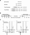
γ-COP binds to p23 but not to Wbp1. (A) Schematic representation of full-length γ-COP and its recombinant trunk and appendage domains. The relative positions of the epitopes of the antibodies against trunk (γ800) and appendage (γ768) domains are indicated by the arrows. (B) Coatomer, full-length γ-COP, γ-trunk, and γ-appendage were incubated with thiopropyl-Sepharose beads coupled to a mutant peptide, p23, or Wbp1 cytoplasmic tail peptides. Input and bound material were analyzed by Western blotting with the anti γ-COP antibodies γ768 (antiappendage) for coatomer, γ-COP, and γ-appendage and γ800 (antitrunk) for γ-trunk.
FIG. 2.
FIG. 2.
Both binding sites for p23 exist in native coatomer. (A) Left: a biotinylated photolabile p23 peptide was used to allow covalent attachment to coatomer. The cross-linked products were purified on streptavidin-coupled beads and analyzed by SDS-polyacrylamide gel electrophoresis with Coomassie blue staining. Right: as a comparison, immunopurified coatomer was loaded on the same gel. The labeled bands were identified by matrix-assisted laser desorption ionization analysis. Note the striking enrichment of γ-COP in the purified cross-linked sample, indicating the specificity of the cross-link. (B) A biotinylated photolabile p23 peptide was used to allow covalent attachment to coatomer. After hydroxylamine cleavage (1) of an asparaginyl-glycine bond present in γ-COP, fragments cross-linked to p23 were purified through streptavidin-coupled beads. There is a unique Asn/Gly bond within γ-COP located at positions 549/550 connecting γ-COP's trunk and appendage domains. Lanes: 3, 7, and 11, mock-treated samples; 4, 8, and 12, hydroxylamine-treated samples; 1, 2, 5, 6, 9, and 10, samples corresponding to a control experiment, which was performed with a nonbiotinylated peptide. In these samples, no unspecific signal can be detected. In lane 12, the black squares indicate the three prominent bands that can be decorated with streptavidin-HRP and that migrate at the expected sizes for full-length γ-COP, γ-trunk, and γ-appendage. In lanes 4 and 8, arrows indicate the γ-appendage and the γ-trunk, respectively. In these two lanes, signal quantification indicates a similar ratio (of 60/40) between recovered noncleaved γ-COP and cleaved γ-COP, indicating that the photoactivable peptide was cross-linked to similar extents to both γ-COP subdomains. Numbers on the left sides of the gels indicate molecular masses.
FIG. 3.
FIG. 3.
The γ-appendage binds to dimers of p23 tails but not to monomers. (A) Structure of the peptides and fusion proteins used in this study. (B) In the binding assay described for Fig. 1, γ-appendage was preincubated with an increasing molar excess of free p23 peptide in solution in either its monomeric or its dimeric form as indicated. The material bound to the beads was analyzed by Western blotting. (C) Chromatograms of gel filtration experiments. γ-Appendage was incubated with a monomeric p23 cytoplasmic tail peptide (left) or with the dimeric form (right). The samples were loaded on a Superdex 200 gel filtration column (Amersham Biosciences), and 50-μl fractions were collected. The fractions were analyzed by Western blotting with antibodies against γ-appendage or p23. Void volume (Vo) and inclusion volume (Vi) are indicated. OD214, optical density at 214 nm.
FIG. 4.
FIG. 4.
The γ-trunk binds to dimers of p23 tails but not to monomers. (A) In the binding assay described for Fig. 1, γ-trunk was preincubated with an increasing molar excess of free p23 peptide in solution either in its monomeric form or in its dimeric form as indicated. The material bound to the beads was analyzed by Western blotting. (B) Five picomoles of γ-appendage was incubated for 1 h at room temperature with no peptide, 50 μM of monomeric p23 peptide, or 50 μM of dimeric p23 peptide as indicated. The samples were then centrifuged for 30 min at 100,000 × g. The pellets (P) and the supernatants (S) were analyzed by Western blotting. The lines indicate that relevant lanes were digitally isolated from the original picture. (C) The experiment was performed as for panel B, except that the γ-trunk was used. In addition, a gel system that allowed detection of the p23 peptides was used.
FIG. 5.
FIG. 5.
Analysis of the binding of dimeric tails of p24 proteins to coatomer. (A) Binding levels of dimeric fusion proteins harboring the cytoplasmic tails of p23, p24, p25, p26, p27, and ERGIC-53 were tested in the microplate assay for the γ-appendage, γ-trunk, full-length γ-COP, and complete coatomer. The KD values derived from the fits are plotted as a histogram. Error bars represent the standard errors of the mean (SEM) calculated from at least three independent experiments. (B) KD values and corresponding Hill coefficients calculated for the interactions of the indicated tails with coatomer. The standard error for each value is indicated in parentheses.
FIG. 6.
FIG. 6.
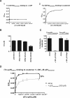
Binding of monomeric cytoplasmic tails to coatomer. (A) Samples of γ-COP (1) or coatomer (2) were analyzed in the microplate-based assay for their binding to (Trx-OST48)monomer. The corresponding data were plotted with the software PRISM (Graphpad), and when possible the data were fitted to a hyperbole. OD405, optical density at 405 nm. (B) Binding to coatomer of monomeric fusion proteins harboring the cytoplasmic tails of p25, Wbp1, DPM2, or OST48 were tested in the microplate-based assay. The KD values derived from the fits are plotted as a histogram. (C) (Trx-p25)monomer or (Trx-p27)dimer (2 μM) was incubated in wells precoated with coatomer. Where indicated, the incubation was performed in the presence of 200 μM of Wbp1 tail peptide. The resulting binding of the fusion proteins was detected with S protein conjugated to HRP. (D) The binding (B/Bmax) of (Trx-p25)dimer to coatomer in the absence (black squares) or in the presence (gray triangles) of 200 μM of p25monomer tail peptide was analyzed in the microplate-based assay. The corresponding data were normalized (the calculated saturation in the absence of competing peptide was set to 1 [Bmax]). As indicated, the plateau in the presence of the competing peptide was decreased by 28%. Error bars represent the SEM calculated from at least three independent experiments.
FIG. 7.
FIG. 7.
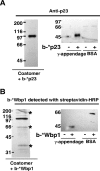
The photoactivable peptide b-*Wbp1 is not as specific as b-*p23. (A) Left: Coatomer at 0.1 μM was incubated with 50 μM biotinylated *p23 photoactivable peptide. After UV-induced cross-linking, the sample was immunoprecipitated with an antibody directed against native coatomer. The sample was then eluted and dissociated with 8 M urea. It was then diluted and incubated with streptavidin-coupled beads in order to purify the cross-linked products. The streptavidin-bound material was then analyzed by Western blotting with an antibody against p23. Right: BSA or γ-appendage at 0.5 μM was mixed with 50 μM of *p23 photoactivable peptide and, after UV-induced cross-linking, was analyzed by Western blotting with an antibody against p23. (B) The experiment was performed as described for panel A except that the biotinylated *Wbp1 photoactivable peptide (b-*Wbp1) was used. The asterisks mark the bands that are probably α-, δ-, and ɛ-COP (from top to bottom). Note that in contrast to b-*p23, b-*Wbp1 showed poor specificity, as it could be cross-linked to multiple coatomer subunits, to the γ-appendage domain, and to BSA. Numbers on the left sides of the gels indicate molecular masses.
FIG. 8.
FIG. 8.
Clustering of p23 tails induces increased avidity for coatomer. Coatomer was analyzed in the microplate-based assay for its binding to (Trx-p23)dimer or to (Trx-p23)tetramer. The corresponding data were plotted with the software PRISM (Graphpad) and were fitted to a hyperbole. Error bars represent the SEM calculated from at least three independent experiments. OD450, optical density at 450 nm.
FIG. 9.
FIG. 9.
A model for the sorting of p24 proteins into COPI vesicles. p23 dimers can recruit Arf-GDP (blue disk) to the membrane (1), allowing nucleotide exchange (2). Once bound to GTP, Arf-GTP (blue square) dissociates from the p23 dimer (3). Coatomer can simultaneously bind to Arf-GTP and the dimer of p23 (4). This results in the stable association of coatomer to membranes (5) and allows the coat to polymerize to build a lattice and eventually form a vesicle. Note that if GTP was replaced by a nonhydrolyzable analog, all the steps would be unchanged.

Similar articles

Cited by

References

    1. Bornstein, P., and G. Balian. 1977. Cleavage at Asn-Gly bonds with hydroxylamine. Methods Enzymol. 47:132-145. - PubMed
    1. Bremser, M., W. Nickel, M. Schweikert, M. Ravazzola, M. Amherdt, C. A. Hughes, T. H. Sollner, J. E. Rothman, and F. T. Wieland. 1999. Coupling of coat assembly and vesicle budding to packaging of putative cargo receptors. Cell 96:495-506. - PubMed
    1. Contreras, I., E. Ortiz-Zapater, and F. Aniento. 2004. Sorting signals in the cytosolic tail of membrane proteins involved in the interaction with plant ARF1 and coatomer. Plant J. 38:685-698. - PubMed
    1. Cosson, P., and F. Letourneur. 1994. Coatomer interaction with di-lysine endoplasmic reticulum retention motifs. Science 263:1629-1631. - PubMed
    1. Dominguez, M., K. Dejgaard, J. Fullekrug, S. Dahan, A. Fazel, J. P. Paccaud, D. Y. Thomas, J. J. Bergeron, and T. Nilsson. 1998. gp25L/emp24/p24 protein family members of the cis-Golgi network bind both COP I and II coatomer. J. Cell Biol. 140:751-765. - PMC - PubMed

Publication types

MeSH terms

LinkOut - more resources