. 2006 Nov;26(21):8136-48.
doi: 10.1128/MCB.00257-06. Epub 2006 Aug 28.

Distinct signaling pathways in TRAIL- versus tumor necrosis factor-induced apoptosis

Affiliations

Distinct signaling pathways in TRAIL- versus tumor necrosis factor-induced apoptosis

Zhaoyu Jin et al. Mol Cell Biol. 2006 Nov.

Abstract

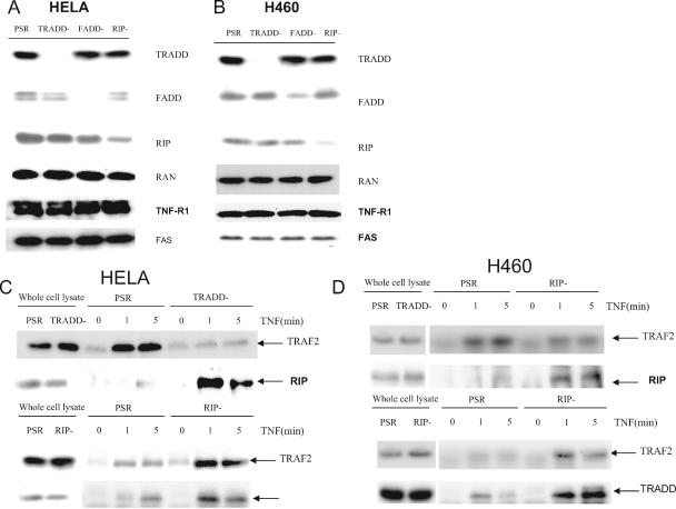
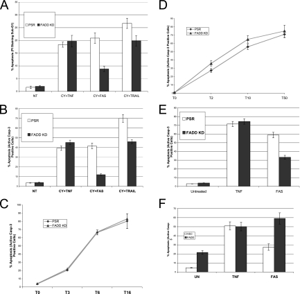
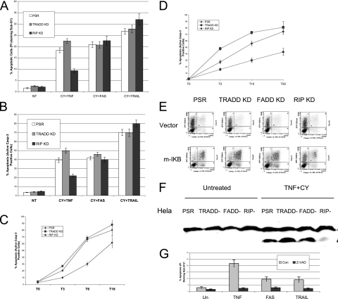
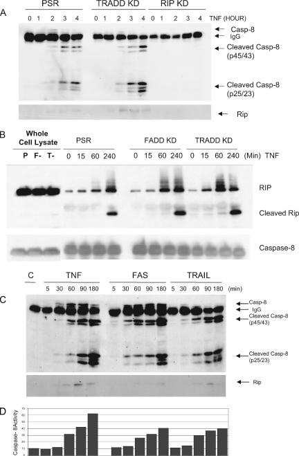
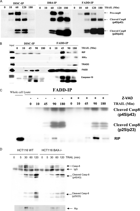
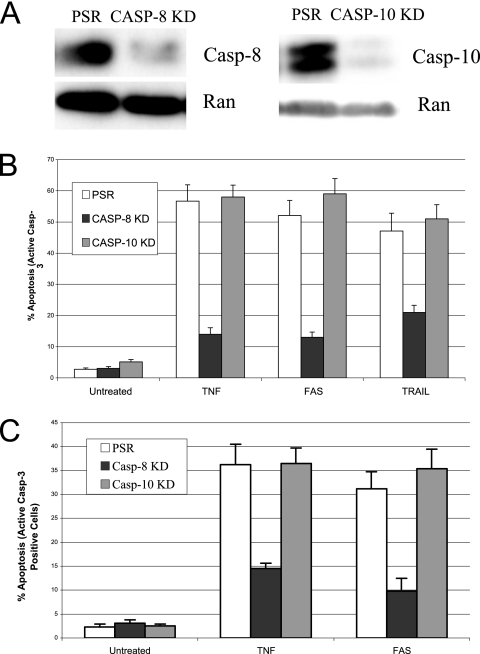
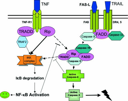
Trimeric tumor necrosis factor (TNF) binding leads to recruitment of TRADD to TNFR1. In current models, TRADD recruits RIP, TRAF2, and FADD to activate NF-kappaB, Jun N-terminal protein kinase (JNK), and apoptosis. Using stable short-hairpin RNA (shRNA) knockdown (KD) cells targeting these adaptors, TNF death-inducing signaling complex immunoprecipitation demonstrates competitive binding of TRADD and RIP to TNFR1, whereas TRAF2 recruitment requires TRADD. Analysis of KD cells indicates that FADD is necessary for Fas-L- or TRAIL- but not TNF-induced apoptosis. Interestingly, TRADD is dispensable, while RIP is required for TNF-induced apoptosis in human tumor cells. TRADD is required for c-Jun phosphorylation upon TNF exposure. RIP KD abrogates formation of complex II following TNF exposure, whereas TRADD KD allows efficient RIP-caspase 8 association. Treatment with TRAIL also induces formation of a complex II containing FADD, RIP, IKKalpha, and caspase 8 and 10, leading to activation of caspase 8. Our data suggest that TNF triggers apoptosis in a manner distinct from that of Fas-L or TRAIL.


Figures

FIG. 1.
FIG. 1.
Recruitment of TRADD, RIP, and TRAF2 to the TNF receptor complex in adaptor KD cells. (A) Whole-cell extracts from stable HeLa KD cell lines carrying nonspecific PSR and specific shRNA for TRADD, FADD, and RIP (PSR, TRADD−, FADD−, RIP−) were prepared and analyzed by Western blotting for TRADD, FADD, RIP, TNFR1, FAS, and Ran (control). (B) Whole-cell extracts from stable H460 knockdown cell lines carrying nonspecific PSR and specific shRNA for TRADD, FADD, RIP (TRADD−, FADD−, RIP−) were prepared and analyzed by Western blotting for TRADD, FADD, RIP, and Ran (control). (C) DISC IP using control (PSR), TRADD knockdown (TRADD−), and RIP knockdown (RIP−) HeLa cells; (D) DISC IP using control (PSR), TRADD knockdown (TRADD−), and RIP knockdown (RIP−) H460 cells. Cells in panel C or D were either untreated or incubated with 500-ng/ml Flag-tagged TNF for 1 or 5 min and then lysed in DISC IP lysis buffer. After normalization of protein content according to the protein assay, cell extracts were immunoprecipitated with anti-flag M2 antibody and protein G-agarose beads. Immunoprecipitates were resolved by SDS-polyacrylamide gel electrophoresis, and Western blotting was performed with anti-TRAF2, anti-RIP, or anti-TRADD, respectively. One percent of the cell extract from each treated sample was used as a control for protein content (input).
FIG. 2.
FIG. 2.
FADD is necessary for Fas-L- and TRAIL-induced apoptosis but not for TNF-induced apoptosis. (A) HeLa control or FADD KD cells were preincubated with cycloheximide (10 μg/ml) and then treated with TNF (20 ng/ml), CH-11 antibody (250 ng/ml), or TRAIL (10 ng/ml) for 4 h, respectively. Apoptosis of HeLa control or FADD KD cells was determined by PI staining and flow cytometry. (B) HeLa control or FADD KD cells were preincubated with cycloheximide (10 μg/ml) and then treated with TNF (20 ng/ml), CH-11 antibody (250 ng/ml), or TRAIL (10 ng/ml) for 4 h, respectively. Apoptosis of HeLa control or FADD KD cells was determined by the active caspase 3 assay. (C) HeLa control or FADD KD cells were preincubated with cycloheximide (10 μg/ml) and then treated with TNF (20 ng/ml) for 3, 6, or 16 h, respectively. Apoptosis was determined by the active caspase 3 assay. (D) HeLa control or FADD KD cells were preincubated with cycloheximide (10 μg/ml) and then treated with 2, 10, or 50 ng/ml TNF for 6 h (T2, T10, and T50, respectively). Apoptosis was determined by the active caspase 3 assay. (E) pSUPER.retro control vector (PSR) or pSR-FADD shRNA (FADD KD) were cotransfected with EGFP at a 10:1 ratio into m-IκB HT-1080 cells. After 48 h, Cells were treated with either TNF (50 ng/ml) or CH-11 (200 ng/ml) for 4 h. Apoptosis was then analyzed by the active caspase 3 assay. (F) pcDNA3.1 control vector (VEC) or pcDNA3.1-FADD (0.2 μg) (FADD) was cotransfected with EGFP at a 10:1 ratio into m-IκB HT-1080 cells. After 48 h, Cells were treated with either TNF (50 ng/ml) or CH-11 (200 ng/ml) for 4 h. Apoptosis was then analyzed by the active caspase 3 assay. The percentage of apoptotic cells from three independent determinations with the corresponding standard deviation is indicated.
FIG. 3.
FIG. 3.
RIP is required for TNF-induced apoptosis but not for Fas-L- or TRAIL-induced apoptosis. (A) HeLa control, TRADD KD, or RIP KD cells were preincubated with cycloheximide (10 μg/ml) and then treated with TNF (20 ng/ml), CH-11 antibody (250 ng/ml), or TRAIL (10 ng/ml) for 4 h, respectively. Apoptosis of HeLa control, TRADD KD, or RIP KD cells was determined by PI staining and flow cytometry. (B) HeLa control, TRADD KD, or RIP KD cells were preincubated with cycloheximide (10 μg/ml) and then treated with TNF (20 ng/ml), CH-11 antibody (250 ng/ml), or TRAIL (10 ng/ml) for 4 h, respectively. Apoptosis of HeLa control, TRADD KD, or RIP KD cells was determined by the active caspase 3 assay. (C) HeLa control, TRADD KD, or RIP KD cells were preincubated with cycloheximide (10 μg/ml) and then treated with TNF (20 ng/ml) for 3, 6, or 16 h (T3, T6, and T16, respectively). Apoptosis was determined by the active caspase 3 assay. (D) HeLa control, TRADD KD, or RIP KD cells were preincubated with cycloheximide (10 μg/ml) and then treated with TNF at 2, 10, or 50 ng/ml for 6 h (T2, T10, and T50, respectively). Apoptosis was determined by the active caspase 3 assay. (E) pcDNA3.1 control vector or pcDNA3.1-m-IκB was cotransfected with EGFP at a 10:1 ratio into HeLa control, TRADD KD, FADD KD, or RIP KD cells. After 48 h, cells were treated with either TNF (20 ng/ml) or CH-11 (100 ng/ml) for 4 h. Apoptosis was then analyzed by the active caspase 3 assay. The percentage of apoptotic cells from three independent determinations with the corresponding standard deviation is indicated. (F) HeLa control, TRADD KD, FADD KD, or RIP KD were preincubated with cycloheximide (10 μg/ml) and then treated with TNF (20 ng/ml) for 4 h. Whole-cell extracts were then prepared and analyzed by Western blotting with PARP antibody. (G) HeLa cells were preincubated with z-VAD-fmk (20 nM) and then treated with TNF (10 ng/ml), CH-11 antibody (100 ng/ml), or TRAIL (10 ng/ml) plus cycloheximide (10 μg/ml) for 4 h. Apoptosis was then analyzed by PI staining and flow cytometry. The percentage of apoptotic cells from three independent determinations with the corresponding standard deviation is indicated.
FIG. 4.
FIG. 4.
RIP is required for the formation of complex II in response to TNF and the assembly of complex II following TNF, FAS-L, or TRAIL treatment; TRADD is not required for TNF-induced apoptosis. (A) HeLa control, TRADD KD, or RIP KD cells were preincubated with cycloheximide (10 μg/ml) and then treated with TNF (20 ng/ml) for different time points as indicated. Analysis of complex II formation was performed by immunoprecipitation using an anti-FADD antibody. Samples were analyzed by Western blotting using antibodies to RIP and caspase 8 (Casp-8). (B) HeLa control, FADD KD, or TRADD KD cells were preincubated with cycloheximide (10 μg/ml) and then treated with TNF (50 ng/ml) for different time points as indicated. A caspase 8 immunoprecipitation was performed using an rabbit anti-caspase 8 antibody, and Western blotting was performed with anti-RIP or anti-caspase 8 antibody. “P” refers to control cells, whereas “F-” and “T-” refer to FADD KD and TRADD KD cells, respectively. (C) I-κB mut HT1080 cells were stimulated with TNF (40 ng/ml), CH-11 (200 ng/ml), or TRAIL (40 ng/ml) for different time points as indicated. Immunoprecipitations were performed using an anti-FADD antibody. Samples were analyzed by Western blotting using antibodies to RIP and caspase 8. (D) A fraction of these cells were incubated with 1 μl of a fluorescein isothiocyanate-IETD-FMK, and the relative caspase 8 activity in these cells was determined by flow cytometry as described in Materials and Methods section.
FIG. 5.
FIG. 5.
The DISC and complex II induced by TRAIL contain different components. H460 cells were stimulated with His-TRAIL (100 ng/ml) for different time points as indicated. Immunoprecipitations were performed using either an anti-His, anti-DR4, or anti-FADD antibody. (A) Samples were analyzed by Western blotting using antibodies to caspase 8 (Casp-8). (B) Samples were analyzed by Western blotting using antibodies to caspase 10, RIP, TRADD, or IKKα. (C) Formation of complex II in Bax−/− HCT-116 cells. H460 cells were treated with TRAIL (100 ng/ml) for different time points as indicated in the presence (+) or absence (−) of z-VAD. Immunoprecipitation was performed by using rabbit anti-FADD antibody. Samples were analyzed by Western blotting using antibodies to Rip and caspase 8. (D) Wild-type (WT) and Bax−/− HCT-116 cells were stimulated with TRAIL (20 ng/ml) for different time points as indicated. Immunoprecipitations were performed using an anti-FADD antibody. Samples were analyzed by Western blotting using antibodies to the indicated proteins.
FIG. 6.
FIG. 6.
Caspase 8, but not caspase 10, is required for death receptor-mediated apoptosis. (A) Whole-cell extracts from HeLa KD cells carrying nonspecific PSR and specific shRNA for caspase 8 and caspase 10 (Casp-8 KD and Casp-10 KD) were prepared and analyzed by Western blotting for caspase 8, caspase 10, and Ran (control). (B) HeLa control or caspase 8 or caspase 10 knockdown cells (PSR, Casp 8 KD, and Casp 10 KD) were preincubated with cycloheximide (10 μg/ml) and then treated with TNF (20 ng/ml), CH-11 antibody (250 ng/ml), or TRAIL (10 ng/ml) for 4 h, respectively. (C) pSUPER.retro control vector (PSR), pRS-casp-8, or pRS-casp-10 was cotransfected with EGFP at a 10:1 ratio into m-IκB HT-1080 cells. After 48 h, cells were treated with either TNF (20 ng/ml) or CH-11 (200 ng/ml) for 6 h. Apoptosis was then analyzed by the active caspase 3 assay. The percentage of apoptotic cells from three independent determinations with the corresponding standard deviation is indicated.
FIG. 7.
FIG. 7.
Model for death receptor-mediated apoptosis. After binding of TNF to TNFR1, RIP and TRADD, followed by TRAF2, are competitively recruited to the membrane receptor complex (complex I). If NF-κB activation triggered by complex I is successful, apoptosis is blocked. Otherwise, TNFR1 dissociates from complex I, and then activated RIP binds to caspase 8/10 and FADD, resulting in the formation of complex II. Activation of caspase 8 in complex II can then lead to apoptosis. Binding of DR4 or FAS to TRAIL or Fas-L recruits FADD to form the DISC. The death ligand then dissociates from the DISC and activated FADD progresses to form the second complex, where caspase 8 is further cleaved and fully activated within complex II. RIP, IKK-alpha, and TRADD are also recruited to complex II, which may be involved in NF-κB activation if apoptosis is blocked.

Similar articles

Cited by

References

    1. Aggarwal, B. B. 2003. Signalling pathways of the TNF superfamily: a double-edged sword. Nat. Rev. Immunol. 3:745-756. - PubMed
    1. Ashkenazi, A., and V. M. Dixit. 1999. Apoptosis control by death and decoy receptors. Curr. Opin. Cell Biol. 11:255-260. - PubMed
    1. Boatright, K. M., M. Renatus, F. L. Scott, S. Sperandio, H. Shin, I. M. Pedersen, J. E. Ricci, W. A. Edris, D. P. Sutherlin, D. R. Green, and G. S. Salvesen. 2003. A unified model for apical caspase activation. Mol. Cell 11:529-541. - PubMed
    1. Brummelkamp, T. R., R. Bernards, and R. Agami. 2002. Stable suppression of tumorigenicity by virus-mediated RNA interference. Cancer Cell 2:243-247. - PubMed
    1. Chen, G., and D. V. Goeddel. 2002. TNF-R1 signaling: a beautiful pathway. Science 296:1634-1635. - PubMed

Publication types

MeSH terms

Substances