

AIDS vaccination studies with an ex vivo feline immunodeficiency virus model: analysis of the accessory ORF-A protein and DNA as protective immunogens
- PMID: 16940498
- PMCID: PMC1563914
- DOI: 10.1128/JVI.00397-06
AIDS vaccination studies with an ex vivo feline immunodeficiency virus model: analysis of the accessory ORF-A protein and DNA as protective immunogens
Abstract
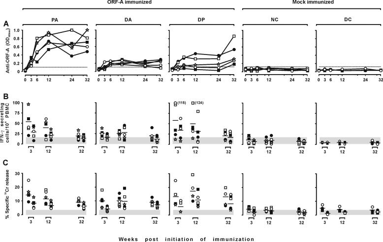
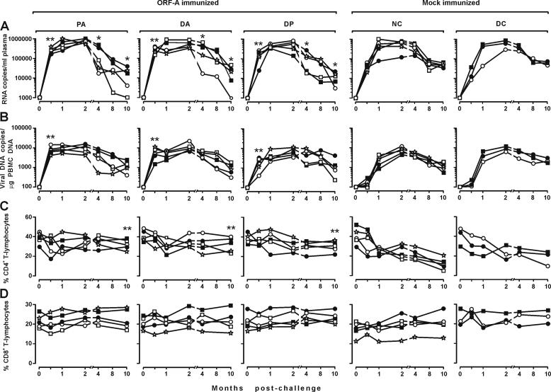
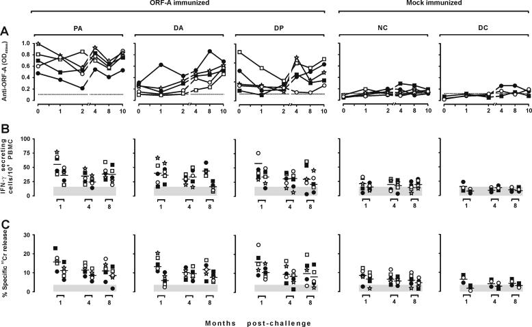
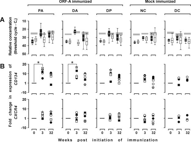
Determining which antigen must be included in AIDS vaccines to confer maximum protection is of utmost importance. In primate models, vaccines consisting of or including accessory viral proteins have yielded conflicting results. We investigated the protective potential of the accessory protein ORF-A of feline immunodeficiency virus (FIV) in cats. All three immunization strategies used (protein alone in alum adjuvant, DNA alone, or DNA prime-protein boost) clearly generated detectable immune responses. Upon challenge with ex vivo homologous FIV, ORF-A-immunized cats showed distinct enhancement of acute-phase infection relative to mock-immunized animals given alum or empty vector DNA. This effect was tentatively attributed to increased expression of the FIV receptor CD134 that was observed in the immunized cats. However, at subsequent sampling points that were continued for up to 10 months postchallenge, the average plasma viral loads of the ORF-A-immunized animals were slightly but consistently reduced relative to those of the control animals. In addition, CD4(+) T lymphocytes in the circulation system declined more slowly in immunized animals than in control animals. These findings support the contention that immunization with lentiviral accessory proteins can improve the host's ability to control virus replication and slow down disease progression but also draw attention to the fact that even simple immunogens that eventually contribute to protective activity can transiently exacerbate subsequent lentiviral infections.
Figures
Similar articles
-
Should accessory proteins be structural components of lentiviral vaccines? Lessons learned from the accessory ORF-A protein of FIV.Vet Immunol Immunopathol. 2008 May 15;123(1-2):144-9. doi: 10.1016/j.vetimm.2008.01.020. Epub 2008 Jan 19. Vet Immunol Immunopathol. 2008. PMID: 18304653 Review.
-
Evaluation of feline immunodeficiency virus ORF-A mutants as candidate attenuated vaccine.Virology. 2005 Feb 20;332(2):676-90. doi: 10.1016/j.virol.2004.12.004. Virology. 2005. PMID: 15680433
-
Gag-specific immune enhancement of lentiviral infection after vaccination with an adenoviral vector in an animal model of AIDS.Vaccine. 2009 Feb 5;27(6):928-39. doi: 10.1016/j.vaccine.2008.11.086. Epub 2008 Dec 12. Vaccine. 2009. PMID: 19070641
-
Structural mapping of CD134 residues critical for interaction with feline immunodeficiency virus.Nat Struct Mol Biol. 2005 Jan;12(1):60-6. doi: 10.1038/nsmb872. Epub 2004 Dec 12. Nat Struct Mol Biol. 2005. PMID: 15592478
-
Tenth anniversary perspectives on AIDS. FIV as a model for AIDS vaccination.AIDS Res Hum Retroviruses. 1994 Mar;10(3):225-8. doi: 10.1089/aid.1994.10.225. AIDS Res Hum Retroviruses. 1994. PMID: 8018382 Review. No abstract available.
Cited by
-
A novel method for producing target cells and assessing cytotoxic T lymphocyte activity in outbred hosts.BMC Biotechnol. 2009 Mar 11;9:18. doi: 10.1186/1472-6750-9-18. BMC Biotechnol. 2009. PMID: 19284578 Free PMC article.
-
Vaccination with vif-deleted feline immunodeficiency virus provirus, GM-CSF, and TNF-alpha plasmids preserves global CD4 T lymphocyte function after challenge with FIV.Vaccine. 2009 Jun 8;27(28):3754-65. doi: 10.1016/j.vaccine.2009.03.081. Epub 2009 Apr 17. Vaccine. 2009. PMID: 19464559 Free PMC article.
-
Streamlined design of a self-inactivating feline immunodeficiency virus vector for transducing ex vivo dendritic cells and T lymphocytes.Genet Vaccines Ther. 2007 Sep 19;5:8. doi: 10.1186/1479-0556-5-8. Genet Vaccines Ther. 2007. PMID: 17880683 Free PMC article.
-
Env-expressing autologous T lymphocytes induce neutralizing antibody and afford marked protection against feline immunodeficiency virus.J Virol. 2010 Apr;84(8):3845-56. doi: 10.1128/JVI.02638-09. Epub 2010 Feb 3. J Virol. 2010. PMID: 20130057 Free PMC article.
-
Feline tetherin efficiently restricts release of feline immunodeficiency virus but not spreading of infection.J Virol. 2011 Jun;85(12):5840-52. doi: 10.1128/JVI.00071-11. Epub 2011 Apr 13. J Virol. 2011. PMID: 21490095 Free PMC article.
References
-
- Allen, T. M., L. Mortara, B. R. Mothé, M. Liebl, P. Jing, B. Calore, M. Piekarczyk, R. Ruddersdorf, D. H. O'Connor, X. Wang, C. Wang, D. B. Allison, J. D. Altman, A. Sette, R. C. Desrosiers, G. Sutter, and D. I. Watkins. 2002. Tat-vaccinated macaques do not control simian immunodeficiency virus SIVmac239 replication. J. Virol. 76:4108-4112. - PMC - PubMed
-
- Amara, R. R., K. Patel, G. Niedziela, P. Nigam, S. Sharma, S. I. Staprans, D. C. Montefiori, L. Chenareddi, J. G. Herndon, H. L. Robinson, H. M. McClure, and F. J. Novembre. 2005. A combination DNA and attenuated simian immunodeficiency virus vaccine strategy provides enhanced protection from simian/human immunodeficiency virus-induced disease. J. Virol. 79:15356-15367. - PMC - PubMed
-
- Broche-Pierre, S., J. Richardson, A. Moraillon, and P. Sonigo. 2005. Evaluation of live feline immunodeficiency virus vaccines with modified antigenic properties. J. Gen. Virol. 86:2495-2506. - PubMed
-
- Burkhard, M. J., and G. A. Dean. 2003. Transmission and immunopathogenesis of FIV in cats as a model for HIV. Curr. HIV Res. 1:15-29. - PubMed
-
- Cafaro, A., A. Caputo, C. Fracasso, M. T. Maggiorella, D. Goletti, S. Baroncelli, M. Pace, L. Sernicola, M. L. Koanga-Mogtomo, M. Betti, A. Borsetti, R. Belli, L. Akerblom, F. Corrias, S. Butto, J. Heeney, P. Verani, F. Titti, and B. Ensoli. 1999. Control of SHIV-89.6P infection of cynomolgus monkeys by HIV-1 Tat protein vaccine. Nat. Med. 5:643-650. - PubMed
Publication types
MeSH terms
Substances
LinkOut - more resources
Full Text Sources
Medical
Research Materials
Miscellaneous
