. 2006 Sep;80(18):8961-9.
doi: 10.1128/JVI.00843-06.

Adeno-associated virus type 2 contains an integrin alpha5beta1 binding domain essential for viral cell entry

Affiliations

Adeno-associated virus type 2 contains an integrin alpha5beta1 binding domain essential for viral cell entry

Aravind Asokan et al. J Virol. 2006 Sep.

Abstract

Integrins have been implicated as coreceptors in the infectious pathways of several nonenveloped viruses. For example, adenoviruses are known to interact with alphaV integrins by virtue of a high-affinity arginine-glycine-aspartate (RGD) domain present in the penton bases of the capsids. In the case of adeno-associated virus type 2 (AAV2), which lacks this RGD motif, integrin alphaVbeta5 has been identified as a coreceptor for cellular entry. However, the molecular determinants of AAV2 capsid-integrin interactions and the potential exploitation of alternative integrins as coreceptors by AAV2 have not been established thus far. In this report, we demonstrate that integrin alpha5beta1 serves as an alternative coreceptor for AAV2 infection in human embryonic kidney 293 cells. Such interactions appear to be mediated by a highly conserved domain that contains an asparagine-glycine-arginine (NGR) motif known to bind alpha5beta1 integrin with moderate affinity. The mutation of this domain reduces transduction efficiency by an order of magnitude relative to that of wild-type AAV2 vectors in vitro and in vivo. Further characterization of mutant and wild-type AAV2 capsids through transduction assays in cell lines lacking specific integrins, cell adhesion studies, and cell surface/solid-phase binding assays confirmed the role of the NGR domain in promoting AAV2-integrin interactions. Molecular modeling studies suggest that NGR residues form a surface loop close to the threefold axis of symmetry adjacent to residues previously implicated in binding heparan sulfate, the primary receptor for AAV2. The aforementioned results suggest that the internalization of AAV2 in 293 cells might follow a "click-to-fit" mechanism that involves the cooperative binding of heparan sulfate and alpha5beta1 integrin by the AAV2 capsids.


Figures

FIG. 1.
FIG. 1.
Anti-integrin antibody-mediated inhibition of AAV2 transduction in 293 cells. Cells grown in 24-well plates (105/well) were incubated for 1 h at 37°C with monoclonal antibodies (20 μg/ml) targeted against different integrin subunits and heterodimers. The cells were then dosed with AAV2-Fluc (MOI, 1,000) in the continued presence of MAbs at 37°C for 24 h prior to the quantitation of luciferase transgene expression in cell lysates. Soluble integrin α5β1 (1 μg/ml) was preincubated with AAV2-Fluc particles for 1 h on ice, and 293 cells were incubated with the mixture for 24 h at 37°C prior to that quantitation of luciferase expression. All experiments were performed in triplicate. Gray bars depict statistically significant data in comparison with the control (P < 0.05). Error bars indicate standard deviations.
FIG. 2.
FIG. 2.
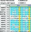
Sequence alignment of the NGR motif found in the 9th type III repeat of fibronectin (25), integrin α5β1 binding NGR domain identified through phage display (23, 24), and a partial section of the VP3 region of AAV serotypes performed in vector NTI. The 511-NGR-513 domain is conserved in the majority of AAV serotypes except AAV4, -5, and -11. AAV4 contains an inverse RGD (DGR) motif, also previously identified as a low-affinity integrin binding motif. Similar residues are depicted with a black font/blue background, dissimilar residues with a black font/white background, and conserved residues in with a red font/yellow background.
FIG. 3.
FIG. 3.
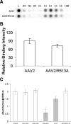
(A) Heparin binding profiles of wt and mut AAV2 particles. Heparin-conjugated agarose beads (500 μl) were loaded in MicroSpin columns, and viral particles (∼1010) were allowed to bind the beads for about 10 min at room temperature. Flowthrough, washes (1× PBS), and eluates at different salt concentrations (in PBS) were collected, and the number of vector genomes in each fraction was determined by dot blot hybridization. Experiments were performed in duplicate. (B) Solid-phase integrin α5β1 binding profiles of wt AAV2 and mut AAV2/R513A capsids. Soluble integrin α5β1 (1 μg/ml) was embedded in nitrocellulose membranes under nondenaturing conditions and probed with either wt or mut AAV2 particles in 5% milk, followed by A20 MAb (1:20 dilution), which recognizes intact AAV2 particles. The extent of binding was quantitated with a horseradish peroxidase-conjugated secondary antibody by using chemiluminescence and densitometric analysis (n = 3; P < 0.05). (C) Competitive inhibition of 293 cell adhesion to fibronectin-coated substrates. Confluent 293 cells were trypsinized and allowed to recover overnight in suspension culture medium. Cells were then incubated with MAbs against integrins α5β1, αVβ3/β5 (20 μg/ml), wt AAV2, or the AAV2/R513A mutant (MOI, ∼108) at 37°C for 1 h to facilitate receptor internalization/blockade. Treated and untreated cells were then allowed to adhere to fibronectin-coated plates for 30 min, fixed, and treated with crystal violet solution (0.5% wt/vol in 20% ethanol in PBS). Cell adhesion was quantitated by determining the UV absorbance of each sample at 595 nm. Gray bars indicate statistically significant data compared with that of the control (n = 4, P < 0.05). Error bars indicate standard deviations.
FIG. 4.
FIG. 4.
Cell surface binding of wt and mutant AAV2 particles. Viral particles were allowed to bind 293 cells for 1 h at 4°C. The cells were then washed three times with 1× PBS, and total cell-associated DNA was extracted using a DNeasy kit. Vector genomes associated with 293 cells were quantitated by dot blot hybridization. All experiments were performed in triplicate. The mutant displayed a statistically significant decrease (P < 0.05) in binding integrin α5β1 and the surface of 293 cells in comparison with that of wt AAV2. Error bars indicate standard deviations.
FIG. 5.
FIG. 5.
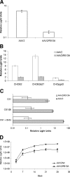
In vitro transduction profiles (luciferase transgene expression) of wt and mut AAV2 vectors (MOI, 1,000) in (A) 293 cells; (B) CHOB2 (integrin α5 negative), CHOB2α27 (integrin α5 transfected), and CHOpgsD (heparan sulfate negative) cells; and (C) CS1 cells before or after treatment with BrdU (2 μM) at 24 h postinfection. Statistical significance was established at P < 0.05 (n = 6). (D) In vivo transduction profiles of wt and mut AAV vectors (dose 1010 vector genomes per mouse) following intramuscular administration into the hind limbs of male BALB/c mice. Images of luciferase transgene expression in vivo were obtained using the Xenogen IVIS 100 imaging system and quantified using Living Image (version 2.5) software. Statistical significance was established with P < 0.05 (n = 4). Error bars indicate standard deviations.
FIG. 6.
FIG. 6.
(A) Three-dimensional model of an AAV2 VP3 trimer viewed down the icosahedral threefold axis of symmetry (wheat, pink, and gray). The location of the NGR integrin recognition sequence (red) is adjacent to heparin binding residues R585/R588 (blue) located within the inner loop. (B) Close-up view of the ribbon structure of the gray and pink monomers with the NGR motif (red) presented as a loop between two beta strands (green). The three-dimensional model of the AAV2 VP3 trimer was generated from VIPER with the available coordinates of AAV2 (PDB accession no. 1LP3) supplied as a template. Subsequent surface rendering was performed using PyMOL.
FIG. 7.
FIG. 7.
Proposed “click-to-fit” model for interaction between the AAV2 capsid and α5β1 integrin. The binding of the native form of wt AAV2 (A) (blue) to cell surface heparan sulfate proteoglycans (HSPG) results in a reversible conformational change (A*) (light blue). The structurally altered virion can then detach and reattach repeatedly to different HSPG on the cell surface until the virion binds both its primary receptor (heparan sulfate) and its secondary receptor, integrin α5β1. Such cooperative interaction of the virion with heparan sulfate and integrin α5β1 results in an irreversible conformation change (B) (dark blue), which in turn could trigger endocytic uptake and subsequent viral entry.

References

    1. Bauer, J. S., J. Varner, C. Schreiner, L. Kornberg, R. Nicholas, and R. L. Juliano. 1993. Functional role of the cytoplasmic domain of the integrin α5 subunit. J. Cell Biol. 122:209-221. - PMC - PubMed
    1. Berns, K. I., and C. Giraud. 1996. Biology of adeno-associated virus. Curr. Top. Microbiol. Immunol. 218:1-23. - PubMed
    1. Bubeck, D., D. J. Filman, and J. M. Hogle. 2005. Cryo-electron microscopy reconstruction of a poliovirus-receptor-membrane complex. Nat. Struct. Mol. Biol. 12:615-618. - PMC - PubMed
    1. Chiu, C. Y., P. Mathias, G. R. Nemerow, and P. L. Stewart. 1999. Structure of adenovirus complexed with its internalization receptor, αvβ5 integrin. J. Virol. 73:6759-6768. - PMC - PubMed
    1. Di Pasquale, G., B. L. Davidson, C. S. Stein, I. Martins, D. Scudiero, A. Monks, and J. A. Chiorini. 2003. Identification of PDGFR as a receptor for AAV-5 transduction. Nat. Med. 9:1306-1312. - PubMed

Publication types

Substances

LinkOut - more resources