. 2006 Sep;80(18):9009-16.
doi: 10.1128/JVI.00969-06.

Mutation at residue 523 creates a second receptor binding site on human parainfluenza virus type 1 hemagglutinin-neuraminidase protein

Affiliations

Mutation at residue 523 creates a second receptor binding site on human parainfluenza virus type 1 hemagglutinin-neuraminidase protein

Tatiana Bousse et al. J Virol. 2006 Sep.

Abstract

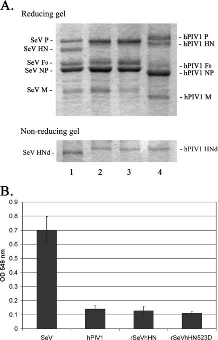
The paramyxovirus hemagglutinin-neuraminidase (HN) is a multifunctional protein mediating hemagglutination (HA), neuraminidase (NA), and fusion promotion activities. It has been a matter of debate whether HN contains combined or separate sites for HA and NA activities. To clear the issue, we determined the presence of the second binding site on human parainfluenza virus (hPIV) type 1, 2, and 3 and Sendai virus (SeV) HN proteins. Results of virus elution from erythrocytes at an elevated temperature and HA inhibition by NA inhibitor BCX-2798 suggest that all hPIVs bind to the receptor only through the NA catalytic site, while SeV HN has an additional receptor binding site. Comparison of SeV and hPIV1 HN sequences revealed two amino acid differences at residues 521 and 523 in the region close to the second binding site identified in Newcastle disease virus HN. We mutated hPIV1 HN at position 523 from Asn to the residue of SeV HN, Asp, and rescued a recombinant SeV that carries the mutated hPIV1 HN by a reverse genetics system. The hPIV1 HN with Asp at position 523 hemagglutinated in the presence of BCX-2798, suggesting that the amino acid difference at position 523 is critical for the formation of a second binding site. Creation of the second binding site on hPIV1 HN, however, did not significantly affect the growth or fusion activity of the recombinant virus. Our study indicates that the presence and requirement of a second binding site vary among paramyxoviruses.


Figures

FIG. 1.
FIG. 1.
Virus adsorption and elution from RBC. Attachment activity of hPIV types 1, 2, and 3 and SeV was analyzed by standard HA assay. Purified viruses (128 HA units) were serially diluted in PBS, and an equal volume of 0.5% human RBC was added to the wells. After incubation for 2 h at 4°C, the plate was then shifted to 34°C and incubated for another 3 h.
FIG. 2.
FIG. 2.
Comparison of the NA activities of parainfluenza viruses. Various amounts of purified viruses were used to analyze viral NA activity by the colorimetric method. All values are averages ± standard deviations (error bars) from three independent experiments. OD 549, optical density at 549 nm.
FIG. 3.
FIG. 3.
Inhibition of NA and HA activities by BCX-2798. (A) NI activities of BCX-2798 against SeV (triangles) and hPIV1 (squares) HN. Purified SeV (0.6 μg) or hPIV1 (5 μg) was preincubated with various concentrations of BCX-2798, and NA activities of the mixture were determined using a colorimetric NA assay. All values are the averages ± standard deviations (error bars) from three independent experiments. (B) HA inhibition of SeV and hPIVs by BCX-2798. Eight HA units of purified viruses were incubated with serially diluted BCX-2798 for 1 h at room temperature. An equal volume of 0.5% human RBC was added to the mixtures and incubated for 1 h at 4°C.
FIG. 4.
FIG. 4.
3D structure of NDV HN showing the location of the second binding site and partial HN amino acid alignment. Sequence comparison between SeV (strain Enders), hPIV1 (strain C35), and NDV (strain Kansas) HNs around the second receptor binding site is shown. Dashes in the hPIV1 HN sequence indicate amino acids identical to those of SeV HN.
FIG. 5.
FIG. 5.
Characterization of rSeVhHN523D. (A) SDS-polyacrylamide gel electrophoresis analysis of purified viruses. Three micrograms of purified SeV (lane 1), rSeVhHN (lane 2), rSeVhHN523D (lane 3), or hPIV1 (lane 4) was fractionated in SDS-10% polyacrylamide gels in reducing and nonreducing conditions. HNd, HN dimer. (B) NA activities of SeV, hPIV1, rSeVhHN, and rSeVhHN523D. Purified viruses (0.4 μg) were used to analyze the NA activity by the colorimetric method. All values are the averages ± standard deviations from three independent experiments. OD 549 nm, optical density at 549 nm.
FIG. 6.
FIG. 6.
Effect of Asn-to-Asp mutation at residue 523 of hPIV1HN on virus attachment activity. (A) An equal amount of purified virus (128 HA units) was serially diluted (1:2 ratio) in PBS and incubated with 0.5% chicken RBC in a 96-well plate for 1 h at 4°C. The plate was then shifted to 34°C and incubated for another 1 h. (B) HI activity of BCX-2798. Eight HA units of purified viruses was incubated with serially diluted BCX-2798 for 1 h at room temperature. An equal volume of 0.5% chicken RBC was added to the mixtures and incubated for another 1 h at 4°C.
FIG. 7.
FIG. 7.
Fusion activity of rSeVhHN and rSeVhHN523D. (A) Syncytium formation of HeLa T4+ cells infected with rSeVhHN or rSeVhHN523D at an MOI of 1. Infected cells were cultured for 24 h and then treated with acetylated trypsin (5 μg/ml) for 30 min and incubated with DMEM containing 10% fetal calf serum for another 4 h. (B) Membrane fusion activity of recombinant SeVs in infected HeLa T4+ cells was determined by content mixing assay. All values are the averages ± standard deviations from three independent experiments.
FIG. 8.
FIG. 8.
Growth kinetics of rSeVhHN (gray triangles) and rSeVhHN523D (black squares) in LLC-MK2 cells. Virus titers were measured at the times indicated on the x axis by plaque assay. The data are means of three experiments with standard errors.

References

    1. Alymova, I. V., G. Taylor, T. Takimoto, T. H. Lin, P. Chand, Y. S. Babu, C. Li, X. Xiong, and A. Portner. 2004. Efficacy of novel hemagglutinin-neuraminidase inhibitors BCX 2798 and BCX 2855 against human parainfluenza viruses in vitro and in vivo. Antimicrob. Agents Chemother. 48:1495-1502. - PMC - PubMed
    1. Aminoff, D. 1961. Methods for the quantitative estimation of N-acetylneuraminic acid and their application to hydrolysates of sialomucoids. Biochem. J. 81:384-392. - PMC - PubMed
    1. Bagai, S., and R. A. Lamb. 1995. Quantitative measurement of paramyxovirus fusion: differences in requirements of glycoproteins between simian virus 5 and human parainfluenza virus 3 or Newcastle disease virus. J. Virol. 69:6712-6719. - PMC - PubMed
    1. Bousse, T., T. Matrosovich, A. Portner, A. Kato, Y. Nagai, and T. Takimoto. 2002. The long noncoding region of the human parainfluenza virus type 1 F gene contributes to the read-through transcription at the M-F gene junction. J. Virol. 76:8244-8251. - PMC - PubMed
    1. Bousse, T., T. Takimoto, W. L. Gorman, T. Takahashi, and A. Portner. 1994. Regions on the hemagglutinin-neuraminidase proteins of human parainfluenza virus type-1 and Sendai virus important for membrane fusion. Virology 204:506-514. - PubMed

Publication types

LinkOut - more resources