. 2006 Sep;80(18):9093-103.
doi: 10.1128/JVI.00895-06.

Alpha2,3 and alpha2,6 N-linked sialic acids facilitate efficient binding and transduction by adeno-associated virus types 1 and 6

Affiliations

Alpha2,3 and alpha2,6 N-linked sialic acids facilitate efficient binding and transduction by adeno-associated virus types 1 and 6

Zhijian Wu et al. J Virol. 2006 Sep.

Abstract

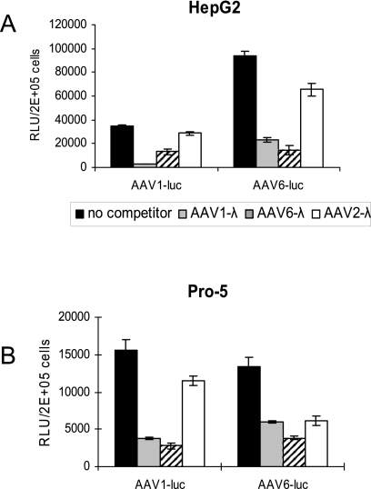
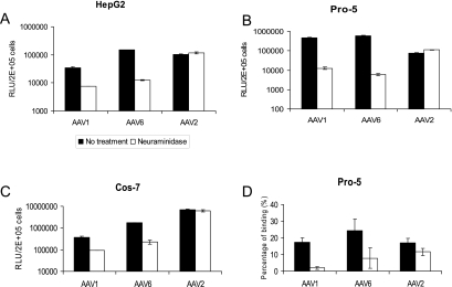
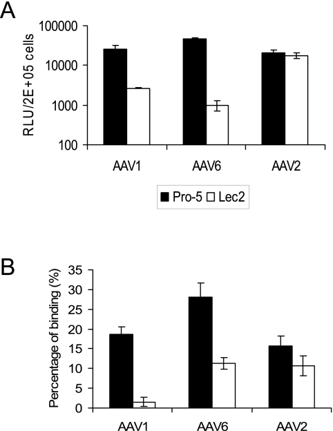
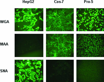
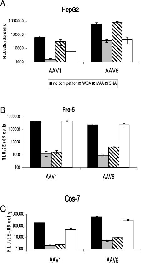
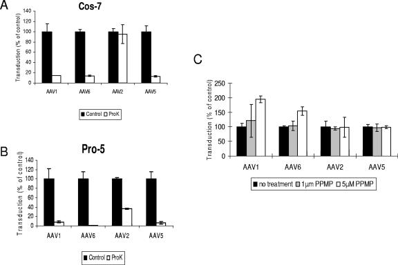
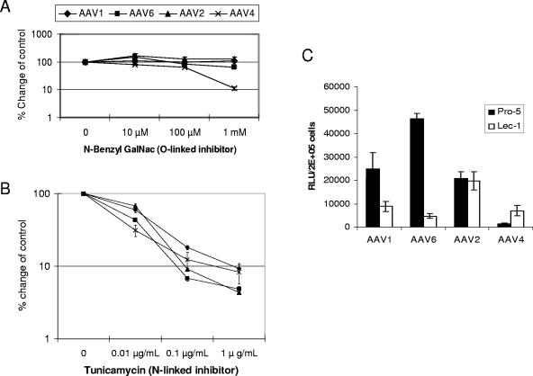
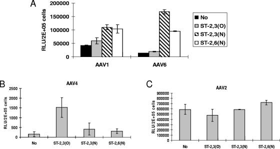
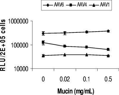
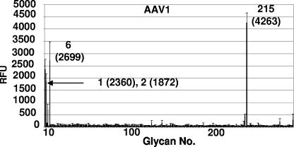
Recombinant adeno-associated viruses (AAVs) are promising vectors in the field of gene therapy. Different AAV serotypes display distinct tissue tropism, believed to be related to the distribution of their receptors on target cells. Of the 11 well-characterized AAV serotypes, heparan sulfate proteoglycan and sialic acid have been suggested to be the attachment receptors for AAV type 2 and types 4 and 5, respectively. In this report, we identify the receptor for the two closely related serotypes, AAV1 and AAV6. First, we demonstrate using coinfection experiments and luciferase reporter analysis that AAV1 and AAV6 compete for similar receptors. Unlike heparin sulfate, enzymatic or genetic removal of sialic acid markedly reduced AAV1 and AAV6 binding and transduction. Further analysis using lectin staining and lectin competition assays identified that AAV1 and AAV6 use either alpha2,3-linked or alpha2,6-linked sialic acid when transducing numerous cell types (HepG2, Pro-5, and Cos-7). Treatment of cells with proteinase K but not glycolipid inhibitor reduced AAV1 and AAV6 infection, supporting the hypothesis that the sialic acid that facilitates infection is associated with glycoproteins rather than glycolipids. In addition, we determined by inhibitor (N-benzyl GalNAc)- and cell line-specific (Lec-1) studies that AAV1 and AAV6 require N-linked and not O-linked sialic acid. Furthermore, a resialylation experiment on a deficient Lec-2 cell line confirmed a 2,3 and 2,6 N-linked sialic acid requirement, while studies of mucin with O-linked sialic acid showed no inhibition effect for AAV1 and AAV6 transduction on Cos-7 cells. Finally, using a glycan array binding assay we determined that AAV1 efficiently binds to NeuAcalpha2-3GalNAcbeta1-4GlcNAc, as well as two glycoproteins with alpha2,3 and alpha2,6 N-linked sialic acids. Taken together, competition, genetic, inhibitor, enzymatic reconstitution, and glycan array experiments support alpha2,3 and alpha2,6 sialic acids that are present on N-linked glycoproteins as primary receptors for efficient AAV1 and AAV6 viral infection.


Figures

FIG. 1.
FIG. 1.
Transduction of cells by luciferase-expressing AAV1 or AAV6 vectors in the presence of competing λ phage DNA-containing AAV1, AAV6, or AAV2 vectors. HepG2 (A) and Pro-5 (B) cells were transduced with a constant amount of AAV1-luc or AAV6-luc vectors in the presence of a 200-fold-excess amount of competing AAV1, AAV6, or AAV2 encapsidated λ phage DNA-containing vector at 37°C for 1 h. The viruses were removed, and the cells were washed three times with medium. The cells were then grown in serum-containing medium. Luciferase expression was tested 24 h posttransduction. RLU, relative light units.
FIG. 2.
FIG. 2.
Neuraminadase treatment of cells reduces AAV1 and AAV6 binding and transduction. (A, B, and C) Transduction assay. Cell surface sialic acid was removed from HepG2 (A), Pro-5 (B), and Cos-7 (C) cells by neuraminadase from Vibrio cholerae prior to transduction with AAV1-luc, AAV6-luc, and AAV2-luc. Twenty-four hours after transduction, cells were analyzed for luciferase expression with a luminometer. RLU, relative light units. (D) Cell binding assay. Pro-5 cells were treated with neuraminidase from Vibrio cholerae or mock treated prior to adding AAV1-luc, AAV6-luc, and AAV2-luc at 4°C. Virus binding was determined by quantitative dot blot hybridization. Values are means from three experiments. Error bars represent standard deviations.
FIG. 3.
FIG. 3.
Transduction and binding on sialic acid-deficient cell lines. (A) Transduction of CHO cells (Pro-5) and sialic acid-deficient CHO cells (Lec-2) with AAV1, AAV6, and AAV2. RLU, relative light units. (B) Binding of AAV1, AAV6, and AAV2 to Pro-5 cells and Lec-2 cells. Values are means from three experiments. Error bars represent standard deviations.
FIG. 4.
FIG. 4.
Lectin binding to HepG2, Cos-7, and Pro-5 cells. Cultures were stained with FITC-labeled lectins that bind to three different carbohydrates as follows: WGA binds sialic acid in any linkage, MAA binds 2,3-linked sialic acid, and SNA binds 2,6-linked sialic acid.
FIG. 5.
FIG. 5.
Lectin competition on HepG2 (A), Pro-5 (B), and Cos-7 (C) cells. Cells growing in 24-well plates (approximately 2 × 10⁵ cells/well) were preincubated at 4°C with 100 μg/ml of each lectin for 10 min. The preincubation solution was removed, and medium containing 100 μg/ml lectin and 2 × 10⁸ virus particles of AAV1-luc, AAV6-luc, or AAV2-luc was added. Cultures were maintained at 4°C for 1 h and then rinsed with medium three times. The cells were grown at 37°C for an additional 24 h in serum-containing media and then assayed for luciferase expression. RLU, relative light units.
FIG. 6.
FIG. 6.
The AAV1 and AAV6 receptor is a glycoprotein rather than a glycolipid. (A and B) Proteinase K treatment reduced AAV1 and AAV6 transduction. Cos-7 (A) and Pro-5 (B) cells were incubated in the presence or absence of 200 μg/ml proteinase K for 1 h at 37°C. The cells were transduced at an MOI of 1 × 10³ for 1 h with AAV1-luc, AAV-6-luc, AAV2-luc, or AAV5-luc, respectively. Luciferase expression was analyzed 24 h after transduction. (C) Glycosphingolipid synthesis inhibitor does not block AAV1 and AAV6 transduction. Cos-7 cells were treated for 40 h with PPMP before transduction with AAV1-luc, AAV6-luc, AAV2-luc, or AAV5-luc. Luciferase expression was analyzed 24 h after transduction.
FIG. 7.
FIG. 7.
N-linked, not O-linked, sialic acid facilitates AAV1 and AAV6 transduction. Cos-7 cells were treated with the indicated doses of N-benzyl GalNAc (A) or tunicamycin (B) for 24 h prior to transduction. Then the cells were transduced with AAV1-luc, AAV6-luc, AAV4-luc, or AAV2-luc. Luciferase expression was analyzed 24 h after transduction. The relative transduction efficiency was determined by comparison with control untreated cells and is presented in log scale. (C) Transduction of CHO cells (Pro-5) and N-linked-glycan-deficient CHO cells (Lec-1) with AAV1-luc, AAV6-luc, AAV2-luc, and AAV4-luc. RLU, relative light units.
FIG. 8.
FIG. 8.
AAV transduction of resialylated sialic acid-deficient Lec-2 cells. The following sialyltransferases were used to add specific sialic acids to the surfaces of Lec-2 cells: α2,3(O)-sialyltransferase, α2,3(N)-sialyltransferase, and α2,6(N)-sialyltransferase. Resialylated Lec-2 cells were then transduced (MOI, 1,000) with either AAV1-luc or AAV6-luc (A), AAV4-luc (B), or AAV2-luc (C), and luciferase expression was measured 24 h posttransduction. RLU, relative light units.
FIG. 9.
FIG. 9.
AAV1 and AAV6 transduction is not inhibited by mucin. Particles (2 × 10⁸) of AAV1-luc, AAV4-luc, or AAV6-luc were incubated with the indicated concentrations of mucin for 30 min at 20°C. Virus alone or virus plus mucin were added to Cos-7 cells growing in 24-well plates (2 × 10⁵ cells/well) in equal volumes of DMEM, and cells were incubated for 1 h at 37°C. Cells were rinsed twice with DMEM and incubated at 37°C. Luciferase expression was tested 24 h later. RLU, relative light units.
FIG. 10.
FIG. 10.
AAV1 capsid glycan specificity on glycan array. A total of 264 glycans were screened for binding to the capsids as described in Materials and Methods. The plot shows the average relative fluorescence (RFU) for the six addresses for each glycan (thick line) versus glycan number. The standard error measurement (SEM) in the fluorescence for the six addresses (thin line) is drawn at the top of each thick line. The four top hits (printed array addresses: 1, AGP; 2, AGP-A; 6, transferrin; 215, NeuAcα2-3GalNAcβ1-4GlcNAcβ) with acceptable SEMs are indicated, with their relative fluorescence levels given in parentheses. All glycans recognized contain a common motif: sialic acid linked to N-acetyl-lactosamine.

References

    1. Bantel-Schaal, U., and H. zur Hausen. 1984. Characterization of the DNA of a defective human parvovirus isolated from a genital site. Virology 134:52-63. - PubMed
    1. Blankinship, M. J., P. Gregorevic, J. M. Allen, S. Q. Harper, H. Harper, C. L. Halbert, D. A. Miller, and J. S. Chamberlain. 2004. Efficient transduction of skeletal muscle using vectors based on adeno-associated virus serotype 6. Mol. Ther. 10:671-678. - PubMed
    1. Blixt, O., S. Head, T. Mondala, C. Scanlan, M. E. Huflejt, R. Alvarez, M. C. Bryan, F. Fazio, D. Calarese, J. Stevens, N. Razi, D. J. Stevens, J. J. Skehel, I. van Die, D. R. Burton, I. A. Wilson, R. Cummings, N. Bovin, C. H. Wong, and J. C. Paulson. 2004. Printed covalent glycan array for ligand profiling of diverse glycan binding proteins. Proc. Natl. Acad. Sci. USA 101:17033-17038. - PMC - PubMed
    1. Bonsdorff, L. V., H. Tolo, E. Lindeberg, T. Nyman, A. Harju, and J. Parkkinen. 2001. Development of a pharmaceutical apotransferrin product for iron binding therapy. Biologicals 29:27-37. - PubMed
    1. Chao, H., Y. Liu, J. Rabinowitz, C. Li, R. J. Samulski, and C. E. Walsh. 2000. Several log increase in therapeutic transgene delivery by distinct adeno-associated viral serotype vectors. Mol. Ther. 2:619-623. - PubMed

Publication types

LinkOut - more resources