. 2006 Sep;80(18):9192-9.
doi: 10.1128/JVI.00555-06.

Inhibition of the type I interferon response by the nucleoprotein of the prototypic arenavirus lymphocytic choriomeningitis virus

Affiliations

Inhibition of the type I interferon response by the nucleoprotein of the prototypic arenavirus lymphocytic choriomeningitis virus

Luis Martínez-Sobrido et al. J Virol. 2006 Sep.

Abstract

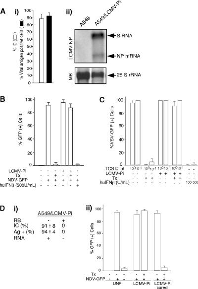
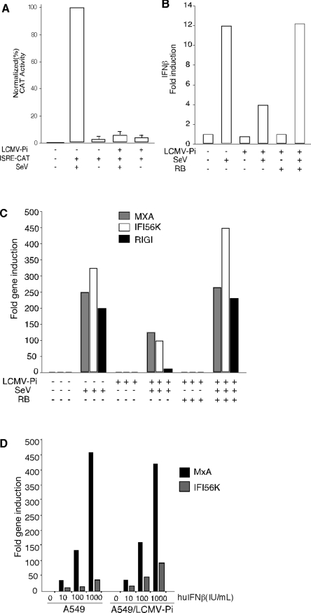
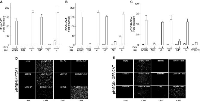
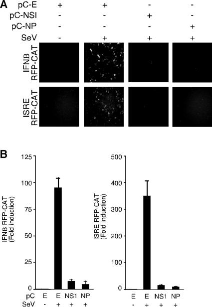
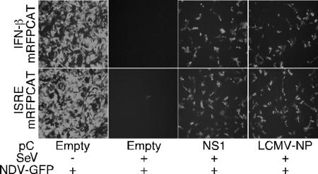
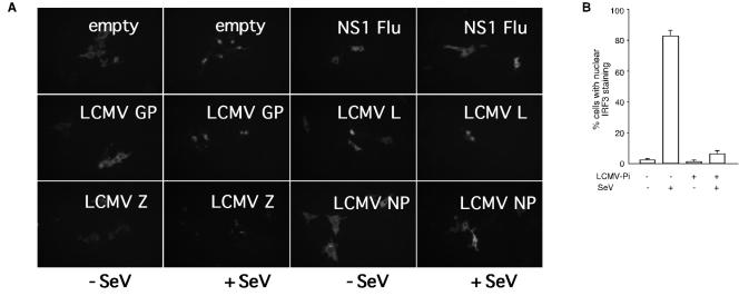
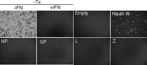
The prototypic arenavirus lymphocytic choriomeningitis virus (LCMV) is a formidable battle horse for the study of viral immunology, as well as viral persistence and associated diseases. Investigations with LCMV have uncovered basic mechanisms by which viruses avoid elimination by the host adaptive immune response. In this study we show that LCMV also disables the host innate defense by interfering with beta interferon (IFN-beta) production in response to different stimuli, including infection with Sendai virus and liposome-mediated DNA transfection. Inhibition of IFN production in LCMV-infected cells was caused by an early block in the IFN regulatory factor 3 (IRF-3) activation pathway. This defect was restored in cells cured of LCMV, indicating that one or more LCMV products are responsible for the inhibition of IRF-3 activation. Using expression plasmids encoding individual LCMV proteins, we found that expression of the LCMV nucleoprotein (NP) was sufficient to inhibit both IFN production and nuclear translocation of IRF-3. To our knowledge, this is the first evidence of an IFN-counteracting viral protein in the Arenaviridae family. Inhibition of IFN production by the arenavirus NP is likely to be a determinant of virulence in vivo.


Figures

FIG. 1.
FIG. 1.
Induction of type I IFN is inhibited in A549/LCMV-Pi cells. (A) Persistence of LCMV in A549 cells. A549 cells were infected with LCMV (MOI of 0.1), and 72 h later, cells were subcultured to establish a persistently infected line (A549/LCMV-Pi). (i) Numbers of cells expressing viral antigen and harboring infectious virus in A549/LCMV-Pi cells were assessed by IF and infectious center assay, respectively. The percentage of infectious centers (IC) was determined as described previously (11). (ii) Levels of viral RNA were determined by Northern blotting using a NP probe to detect S RNA (replication) and NP mRNA (transcription). MB, methylene blue staining of the membrane to detect 28S rRNA. (B) A549 or A549/LCMV-Pi cells were mock transfected (Tx −) or transfected (Tx +) with 2 μg of empty pC plasmid (2, 32). Twenty-four hours posttransfection, cells were infected with NDV-GFP (MOI of 2) (+), and at 24 h p.i., GFP expression was assessed. For a control, A549 cells were treated with 500 IU/ml of human IFN-β (huIFNβ) (+). (C) Vero cells treated (12 h) with TCS from A549 cells or A549/LCMV-Pi cells that had been mock transfected or transfected with empty plasmid. Treated Vero cells were infected with VSV expressing GFP (MOI of 1). TCS from A549 cells treated with human IFN-β were used as controls. Dilut, dilutions. (D) Cells cured of LCMV infection restored their ability to produce type I IFN in response to LF-mediated DNA transfection. (i) Characterization of cells cured of LCMV infection by RB treatment. Ag +, antigen positive. (ii) A549, A549/LCMV-Pi, and RB-treated A549/LCMV-Pi were mock transfected or transfected with empty plasmid. Sixteen hours after transfection, cells were infected with NDV-GFP (MOI of 2), and GFP expression was determined at 24 h p.i. UNF, uninfected.
FIG. 2.
FIG. 2.
SeV-mediated activation of an ISRE promoter, as well as induction of IFN and ISGs, but not the IFN-β induced antiviral state, are inhibited in A549/LCMV-Pi cells. (A) A549 and A549/LCMV-Pi cells were transfected with 1 μg of the ISRE-CAT reporter plasmid (+). After transfection, cells were mock infected (−) or infected with SeV (+). Twenty-four hours later, cell lysates were prepared for CAT assays. (B, C) A549, A549/LCMV-Pi, and RB-cured A549/LCMV-Pi cells were mock infected (−) or infected with SeV (+). At 24 h p.i., total RNA was isolated and quantitative RT-PCR was performed by using specific primers (shown below) to determine the levels of IFN-β mRNA (B), as well as MxA, IFI56K, and RIG-I mRNA (C). (D) Persistence of LCMV in A549 cells does not prevent the type I IFN-induced antiviral state. A549 and A549/LCMV-Pi cells were treated with human IFN-β (huIFNβ) (0, 10, 100, and 1,000 U/ml). Twenty-four hours after IFN treatment, total RNA was isolated and mRNA levels for MxA and IFI56K were determined by qRT-PCR. RT was done using random hexamers as primers. The gene-specific primers used for qPCR were as follows: IFN-β (sense, 5′-GTCAGAGTGGAATCCTAAG-3′; antisense, 5′-ACAGCATCTGCTGGTTGAAG-3′); Mx1 (sense, 5′-CGTGGTGATTTAGCAGGAAG-3′; antisense, 5′-TGCAAGGTGGAGCGATTCTG-3′); RIG-I (sense, 5′-AAAGCCTTGGCATGTTACAC-3′; antisense, 5′-GGCTTGGGATGTGGTCTACT-3′); IFI56K (sense, 5′-TCGGAGAAAGGCATTAGATC-3′; antisense, 5′-GACCTTGTCTCACAGAGTTC-3′); and actin (sense, 5′-ACTGGAACGGTGAAGGTGAC-3′; antisense, 5′-GTGGACTTGGGAGAGGACTG-3′).
FIG. 3.
FIG. 3.
LCMV NP inhibits SeV-mediated activation of IFN-β, ISRE, and IRF promoters. Cells (293T) were cotransfected (+) with 0.5 μg of the different reporter plasmids, together with 4 μg of the indicated LCMV expression plasmids, and 24 h later, cells were mock infected (−) or infected with SeV (+). CAT (A and B), luciferase (C), and GFP (D and E) expression was determined 24 h p.i.
FIG. 4.
FIG. 4.
SeV-mediated activation of IFN-β and ISRE promoters is inhibited in LCMV-NP-transfected cells. 293T cells were transfected as described in the legend to Fig. 3, but using the monomeric red fluorescence protein fused to CAT under the IFN-β (IFN-β mRFP-CAT) or ISRE (ISRE mRFP-CAT) promoter. Twenty-four hours later, cells were mock infected (−) or infected with SeV (+), and 16 h later, activation of the reporter promoters was assessed by epifluorescene and CAT assay. (A) Expression of monomeric RFP was examined by epifluorescence microscopy. (B) Normalized CAT expression levels are shown as changes in induction compared to the values obtained for uninfected cells that were transfected with an empty (E) control plasmid.
FIG. 5.
FIG. 5.
NDV-GFP bioassay. Supernatants from transfected and infected 293T cells shown in Fig. 4 were treated with UV and added to fresh Vero cells. Sixteen hours later, cells were infected with the NDV-GFP (MOI of 2), and at 24 h p.i., GFP expression was detected by epifluorescence.
FIG. 6.
FIG. 6.
Nuclear translocation of IRF-3 is inhibited in both LCMV-PI and LCMV-NP-transfected cells. (A) Vero cells were cotransfected with GFP-tagged IRF-3 (1 μg) together with 4 μg of the indicated LCMV expression plasmids. Twenty-four hours later, cells were mock infected or infected with SeV, and at 16 h p.i. nuclear translocation of GFP-tagged IRF-3 was assessed by epifluorescence. (B) LCMV persistently infected (LCMV-Pi) and control Vero cells were transfected with GFP-tagged IRF-3, and 24 h later, cells were mock infected or infected with SeV. At 16 h p.i. we determined the percentage of cells showing nuclear translocation of GFP-IRF3 in mock-infected or LCMV-Pi Vero cells that were also either mock infected or infected with SeV.
FIG. 7.
FIG. 7.
Expression of LCMV proteins does not prevent the antiviral state induced by interferon. Vero cells were transfected with 2 μg of the indicated expression plasmids, and 24 h later, the cells were treated with human IFN-β (1000 U/ml). Twenty-four hours after IFN treatment, cells were infected with NDV-GFP. A negative control (empty plasmid) and a positive control (Nipah W) were used to validate the assay. Additional controls included Vero cells (top panels) not treated (−IFN) or pretreated (+IFN) with human IFN-β (1,000 U/ml). −Tx, nontransfected cells.

References

    1. Baize, S., J. Kaplon, C. Faure, D. Pannetier, M. C. Georges-Courbot, and V. Deubel. 2004. Lassa virus infection of human dendritic cells and macrophages is productive but fails to activate cells. J. Immunol. 172:2861-2869. - PubMed
    1. Basler, C. F., A. Mikulasova, L. Martinez-Sobrido, J. Paragas, E. Muhlberger, M. Bray, H. D. Klenk, P. Palese, and A. Garcia-Sastre. 2003. The Ebola virus VP35 protein inhibits activation of interferon regulatory factor 3. J. Virol. 77:7945-7956. - PMC - PubMed
    1. Basler, C. F., X. Wang, E. Muhlberger, V. Volchkov, J. Paragas, H. D. Klenk, A. Garcia-Sastre, and P. Palese. 2000. The Ebola virus VP35 protein functions as a type I IFN antagonist. Proc. Natl. Acad. Sci. USA 97:12289-12294. - PMC - PubMed
    1. Beutler, B. 2004. Innate immunity: an overview. Mol. Immunol. 40:845-859. - PubMed
    1. Beyer, W. R., D. Popplau, W. Garten, D. von Laer, and O. Lenz. 2003. Endoproteolytic processing of the lymphocytic choriomeningitis virus glycoprotein by the subtilase SKI-1/S1P. J. Virol. 77:2866-2872. - PMC - PubMed

Publication types

LinkOut - more resources