. 2006 Sep;80(18):9279-87.
doi: 10.1128/JVI.00659-06.

Modulation of the unfolded protein response by the severe acute respiratory syndrome coronavirus spike protein

Affiliations

Modulation of the unfolded protein response by the severe acute respiratory syndrome coronavirus spike protein

Ching-Ping Chan et al. J Virol. 2006 Sep.

Abstract

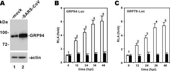
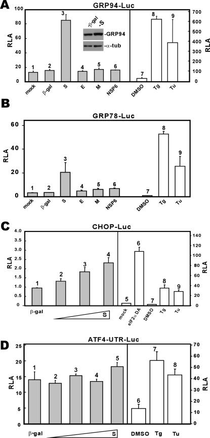
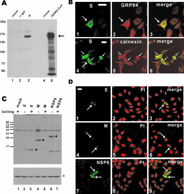
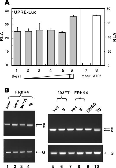
Perturbation of the function of endoplasmic reticulum (ER) causes stress leading to the activation of cell signaling pathways known as the unfolded protein response (UPR). Severe acute respiratory syndrome (SARS) coronavirus (SARS-CoV) uses ER as a site for synthesis and processing of viral proteins. In this report, we demonstrate that infection with SARS-CoV induces the UPR in cultured cells. A comparison with M, E, and NSP6 proteins indicates that SARS-CoV spike (S) protein sufficiently induces transcriptional activation of several UPR effectors, including glucose-regulated protein 78 (GRP78), GRP94, and C/EBP homologous protein. A substantial amount of S protein accumulates in the ER. The expression of S protein exerts different effects on the three major signaling pathways of the UPR. Particularly, it induces GRP78/94 through PKR-like ER kinase but has no influence on activating transcription factor 6 or X box-binding protein 1. Taken together, our findings suggest that SARS-CoV S protein specifically modulates the UPR to facilitate viral replication.


Figures

FIG. 1.
FIG. 1.
Infection with SARS-CoV induces GRP94 and GRP78. (A) Induction of GRP94 expression. FRhK4 cells were either mock infected or infected with SARS-CoV. Cells harvested at 24 hpi were lysed and immunoblotted with anti-GRP94 and anti-β-actin. (B and C) Transcriptional activation of GRP94 and GRP78 promoters. Vero cells were transfected with pGRP94/78-Luc plasmid at 24 h before infection with SARS-CoV (□) or with UVC-inactivated SARS-CoV (▪). Cells were harvested for dual luciferase assay at the indicated time points. RLA values shown represent the means ± standard deviations of the results from three separate infections. Control plasmid pRLSV40 expressing Renilla luciferase was cotransfected into cells and used for normalization of transfection efficiency.
FIG. 2.
FIG. 2.
Influence of SARS-CoV proteins on the UPR. (A and B) SARS-CoV S protein activates GRP94 and GRP78 promoters. 293FT cells were transiently cotransfected with pGRP78/94-Luc plus a pLenti-based expression vector for the indicated protein. Control cells transfected with pGRP78/94-Luc alone were treated with dimethyl sulfoxide (DMSO), Tg (300 nM), or Tu (5 μg/ml) for 16 h. Cells were harvested 48 hpi for dual luciferase assay. Expression levels of GRP94 and α-tubulin (α-tub) in β-galactosidase (β-gal)- and S-expressing cells were verified by Western blotting (inset). (C) SARS-CoV S protein modestly activates CHOP promoter. 293FT cells were cotransfected with pCHOP-Luc and escalating amounts of pLenti-S. Cells were harvested for dual luciferase assay as described for panel A. A DA form of eIF2α was used as a positive control in this analysis, as were Tg (300 nM) and Tu (5 μg/ml). Transcription from the CHOP promoter is significantly induced by eIF2α DA, Tg, and Tu. (D) SARS-CoV S protein does not significantly affect ATF4 translation. 293FT cells were cotransfected with pATF4-UTR-Luc and escalating amounts of pLenti-S. Cells were harvested for dual luciferase assay as described for panel A. Tg (300 nM) and Tu (5 μg/ml) were used as positive controls in this analysis. pATF4-UTR-Luc contains the 5′ UTR of human ATF4 fused to the coding region of firefly luciferase. This construct is similar to those described for mouse ATF4 (32, 52).
FIG. 3.
FIG. 3.
Expression of SARS-CoV proteins in cultured cells. (A) Western blot analysis of S protein expression. pLenti-β-gal-transfected and pLenti-S-transfected 293FT cells (lanes 2 and 3) and SARS-CoV-infected FRhk4 cells (lane 5) were lysed and immunoblotted with rabbit polyclonal anti-S antibody. The arrow highlights S protein. (B) Subcellular localization of SARS-CoV S protein. HeLa cells were transfected with pLenti-S and then costained with rabbit anti-V5 and either rat anti-GRP94 (panel 2) or mouse anti-calnexin (panel 5). The S and GRP94-calnexin fluorescent signals are shown in merged images, and colocalization is shown in yellow (panels 3 and 6). Transfected cells are highlighted with arrows. Bar, 30 μm. (C and D) Expression of other SARS-CoV proteins. 293FT cells were transfected with pLenti-E/M/NSP6, and protein expression was analyzed by immunoblotting (C) or confocal immunostaining (D) with rabbit polyclonal anti-V5 antibody. To prevent thermal aggregation of M and NSP6 (28), protein samples were not heated before being loaded onto the SDS-polyacrylamide gel electrophoresis gel. Nuclear morphology of cells was visualized by propidium iodide (PI) staining. In panel C, the arrowheads highlight E/M/NSP6 proteins and the asterisk indicates the control protein α-tubulin.
FIG. 4.
FIG. 4.
Activation of GRP94/GRP78 by SARS-CoV S protein requires PERK and eIF2α phosphorylation. 293FT cells were cotransfected with pGRP78/94-Luc and expression vectors for the indicated combinations of proteins. Cells were harvested for dual luciferase assay as described for Fig. 2 (A to D). The steady-state levels of phosphorylated (*) and total (#) eIF2α in cells treated with DMSO (lanes 1 and 7) or Tg (300 nM; lanes 2 and 8), mock infected (lanes 3 and 9) or infected with SARS-CoV (lanes 4 and 10), and transfected with pLenti-Topo empty vector alone (lanes 5 and 11) or with pLenti-S (lanes 6 and 12) were verified by Western blotting (E). Treatment with Tg was used as a positive control in this experiment. WT, wild type.
FIG. 5.
FIG. 5.
SARS-CoV S protein does not stimulate UPRE or XBP1 splicing. (A) 293FT cells were cotransfected with pUPRE-Luc and escalating amounts of pLenti-S. In the control group, an expression plasmid for a constitutively active version of ATF6 containing 1 to 373 amino acids was cotransfected. Cells were harvested for dual luciferase assay as described for Fig. 2. ATF6 (1-373) is known to stimulate UPRE, as reported previously (8). (B) 293FT and FRhK4 cells were mock infected (lane 1), infected with SARS-CoV (lane 2), transfected with pLenti-Topo empty vector (vec; lanes 5 and 7), transfected with pLenti-S plasmid (lanes 6 and 8), or treated with MG132 (lane 3), Tg (lanes 4 and 10), or DMSO (lane 9) for 8 h. Unspliced (u) and spliced (s) forms of XBP1 transcript were examined by reverse transcription-PCR as described previously (26). GAPDH mRNA (G) was also detected as a positive control.
FIG. 6.
FIG. 6.
SARS-CoV S protein does not stimulate ATF6-dependent transcriptional activity. 293FT cells were cotransfected with pGal-Luc, pGal-ATF6, and escalating amounts of pLenti-S. Cells were harvested for dual luciferase assay as described for Fig. 2. Tg (300 nM) and Tu (5 μg/ml) were used as positive controls in this analysis.

References

    1. Ball, J. M., T. Peng, C. Q. Y. Zeng, A. P. Morris, and M. K. Estes. 1996. Age-dependent diarrhea induced by a rotaviral nonstructural glycoprotein. Science 272:101-104. - PubMed
    1. Baumeister, P., S. Luo, W. C. Skarnes, G. Sui, E. Seto, Y. Shi, and A. S. Lee. 2005. Endoplasmic reticulum stress induction of the Grp78/BiP promoter: activating mechanisms mediated by YY1 and its interactive chromatin modifiers. Mol. Cell. Biol. 25:4529-4540. - PMC - PubMed
    1. Benali-Furet, N. L., M. Chami, L. Houel, F. De Giorgi, F. Vernejoul, D. Lagorce, L. Buscail, R. Bartenschlager, F. Ichas, R. Rizzuto, and P. Paterlini-Bréchot. 2005. Hepatitis C virus core triggers apoptosis in liver cells by inducing ER stress and ER calcium depletion. Oncogene 24:4921-4933. - PubMed
    1. Boyce, M., K. F. Bryant, C. Jousse, K. Long, H. P. Harding, D. Scheuner, R. J. Kaufman, D. Ma, D. M. Coen, D. Ron, and J. Yuan. 2005. A selective inhibitor of eIF2α dephosphorylation protects cells from ER stress. Science 307:935-939. - PubMed
    1. Boyce, M., and J. Yuan. 2006. Cellular response to endoplasmic reticulum stress: a matter of life or death. Cell Death Differ. 13:363-373. - PubMed

Publication types

MeSH terms