. 2006 Sep;80(18):9310-21.
doi: 10.1128/JVI.02615-05.

Herpes simplex virus type 1 latently infected neurons differentially express latency-associated and ICP0 transcripts

Affiliations

Herpes simplex virus type 1 latently infected neurons differentially express latency-associated and ICP0 transcripts

Séverine Maillet et al. J Virol. 2006 Sep.

Abstract

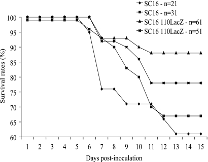
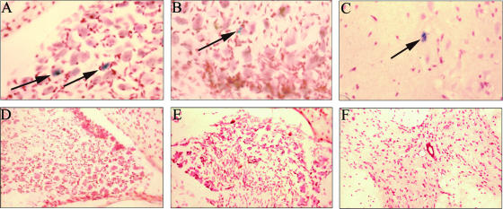
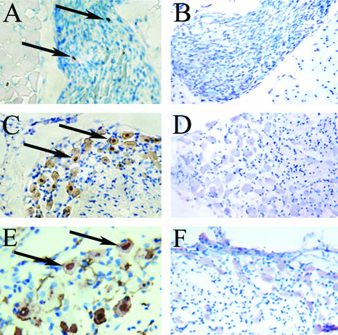
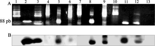
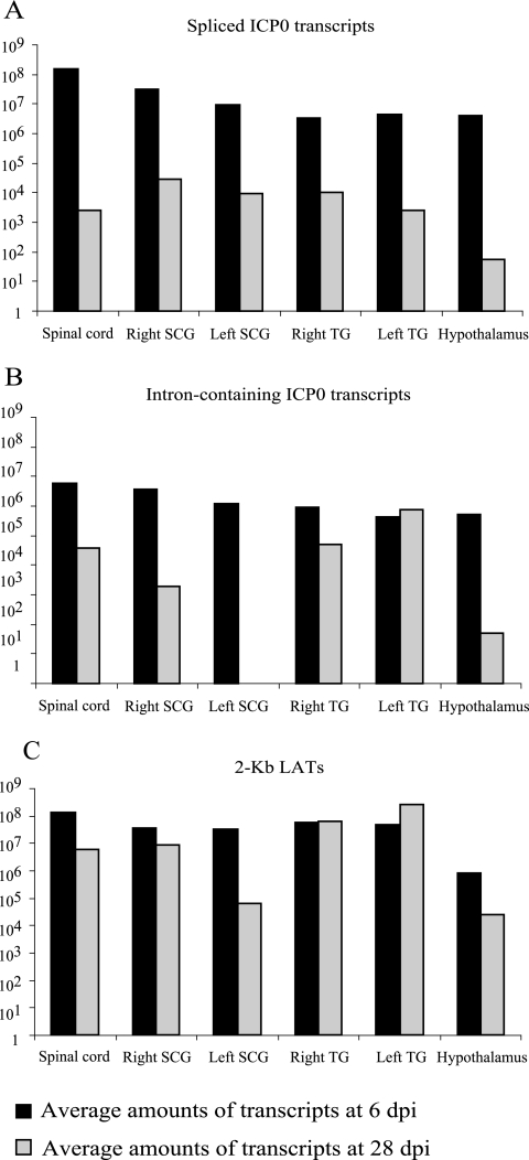
During the latent phase of herpes simplex virus type 1 (HSV-1) infection, the latency-associated transcripts (LATs) are the most abundant viral transcripts present in neurons, but some immediate-early viral transcripts, such as those encoding ICP0, have also been reported to be transcribed in latently infected mouse trigeminal ganglia (TG). A murine oro-ocular model of herpetic infection was used to study ICP0 gene expression in the major anatomical sites of HSV-1 latency, including the TG, superior cervical ganglion, spinal cord, and hypothalamus. An HSV-1 recombinant strain, SC16 110LacZ, revealed ICP0 promoter activity in several neurons in latently infected ganglia, and following infection with wild-type HSV-1 strain SC16, in situ hybridization analyses identified ICP0 transcripts in the nuclei of neurons at times consistent with the establishment of latency. Reverse transcription (RT)-PCR assays performed on RNA extracted from latently infected tissues indicated that ICP0 transcripts were detected in all anatomical sites of viral latency. Furthermore, quantitative real-time RT-PCR showed that neurons differentially expressed the LATs and ICP0 transcripts, with splicing of ICP0 transcripts being dependent on the anatomical location of latency. Finally, TG neurons were characterized by high-level expression of LATs and detection of abundant unspliced ICP0 transcripts, a pattern markedly different from those of other anatomical sites of HSV-1 latency. These results suggest that LATs might be involved in the maintenance of HSV-1 latency through the posttranscriptional regulation of ICP0 in order to inhibit expression of this potent activator of gene expression during latency.


Figures

FIG. 1.
FIG. 1.
Survival rates of mice infected with either the recombinant SC16 110LacZ or the wild-type SC16 strain of HSV-1. Four independent experiments were performed: 21 and 31 mice were inoculated with 1 μl of a suspension containing 106 PFU of SC16, and 61 and 51 mice were infected with SC16 110LacZ.
FIG. 2.
FIG. 2.
Detection of β-galactosidase activity on latently infected mice. Animals were infected with SC16 110LacZ (A, B, and C) or mock infected (D, E, and F) and sacrificed at 28 dpi. ICP0 promoter activity was detected using the β-galactosidase detection assay (blue staining within the cytoplasms). (A and D) SCG; (B and E) TG; (C and F) spinal cord; (A) two positive cells in the superior cervical ganglion (arrows); (B) one positive cell in the trigeminal ganglion (arrow); (C) one positive cell in the spinal cord (arrow).
FIG. 3.
FIG. 3.
Detection of ICP0 transcripts by ISH. SC16 (A, C, and E)- or mock (B, D, and F)-infected mice were sacrificed when latency was established at 28 dpi. Cells positive for ICP0 transcripts, revealed by dark brown staining, are located predominantly within the nuclei. (A and B) SCG at 28 dpi; (A) two positive cells; (C and D) TG at 28 dpi; (C) numerous positive cells; (E and F) TG at 90 dpi; (E) numerous positive cells. Arrows indicate the nuclei of positive cells.
FIG. 4.
FIG. 4.
Amplification of ICP0 transcripts using RT-PCR. (A) RT-PCR products were separated on an agarose gel. (B) Southern blot of the amplification products hybridized with the internal probe O3. Lanes 1, 2, and 3, spinal cord; lanes 4, 5 and 6, SCG; lanes 7, 8 and 9, TG; lanes 10, 11, and 12, hypothalamus; lane 13, negative control without the reverse transcriptase step; lanes 1, 4, 7, and 10, mock-infected mice; lanes 2, 5, 8, and 11, infected mice sacrificed at 6 dpi (acute phase); lanes 3, 6, 9, and 12, infected mice sacrificed at 28 dpi (latent phase of infection). An 88-base pair amplified fragment was observed for acutely and latently infected tissues (middle and right wells) but not for mock-infected tissues. The signal specificity was confirmed by Southern blotting.
FIG. 5.
FIG. 5.
Bar graphs comparing HSV-1 transcripts at 6 dpi and 28 dpi. (A) Spliced ICP0 transcripts; (B) intron-containing ICP0 transcripts; (C) 2-kb LAT transcripts.
FIG. 6.
FIG. 6.
Amplification of TK and UL18 transcripts, using RT-PCR from spinal cord. RNA extracted from mock (lane 1)-, latently (lanes 2 to 5), and acutely (lanes 6 to 8) infected tissues, corresponding to mice L10 to L13 and A2 to A4, respectively. Panels A and B show agarose gels for TK and UL18 amplification products, respectively. No amplification product could be detected in mock- and latently infected RNA extracts. (C) Southern blot using a digoxigenin-labeled probe specific to the UL18 amplification product. Equivalent results were obtained for UL18 transcripts in TG, SCG, and hypothalamus and for TK transcripts in TG, SCG, spinal cord, and hypothalamus. Panels D and E show semiquantitative evaluations of the RT-PCR sensitivity for TK and UL18 transcripts, respectively, using increasing amounts of corresponding transcripts (from wells left to right, 0 to 106 copies).

References

    1. Alvira, M. R., W. F. Goins, J. B. Cohen, and J. C. Glorioso. 1999. Genetic studies exposing the splicing events involved in herpes simplex virus type 1 latency-associated transcript production during lytic and latent infection. J. Virol. 73:3866-3876. - PMC - PubMed
    1. Arthur, J., S. Efstathiou, and A. Simmons. 1993. Intranuclear foci containing low abundance herpes simplex virus latency-associated transcripts visualized by non-isotopic in situ hybridization. J. Gen. Virol. 74:1363-1370. - PubMed
    1. Block, T. M., and J. M. Hill. 1997. The latency associated transcripts (LAT) of herpes simplex virus: still no end in sight. J. Neurovirol. 3:313-321. - PubMed
    1. Bruni, R., and B. Roizman. 1996. Open reading frame P—a herpes simplex virus gene repressed during productive infection encodes a protein that binds a splicing factor and reduces synthesis of viral proteins made from spliced mRNA. Proc. Natl. Acad. Sci. USA 93:10423-10427. - PMC - PubMed
    1. Burton, E. A., C. S. Hong, and J. C. Glorioso. 2003. The stable 2.0-kilobase intron of the herpes simplex virus type 1 latency-associated transcript does not function as an antisense repressor of ICP0 in nonneuronal cells. J. Virol. 77:3516-3530. - PMC - PubMed

Publication types

Substances

LinkOut - more resources