

Clinical-histopathological correlation in a case of Coats' disease
- PMID: 16942617
- PMCID: PMC1564043
- DOI: 10.1186/1746-1596-1-24
Clinical-histopathological correlation in a case of Coats' disease
Abstract
Background: Coats' disease is a non-hereditary ocular disease, with no systemic manifestation, first described by Coats in 1908. It occurs more commonly in children and has a clear male predominance. Most patients present clinically with unilateral decreased vision, strabismus or leukocoria. The most important differential diagnosis is unilateral retinoblastoma, which occurs in the same age group and has some overlapping clinical manifestations.
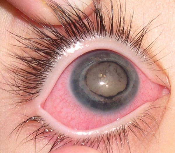
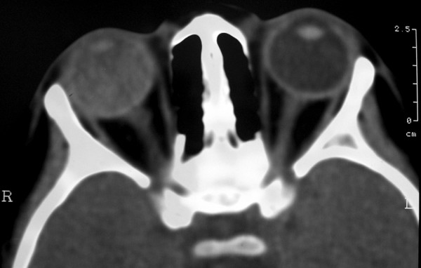
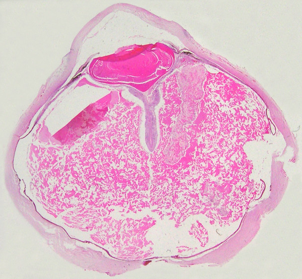
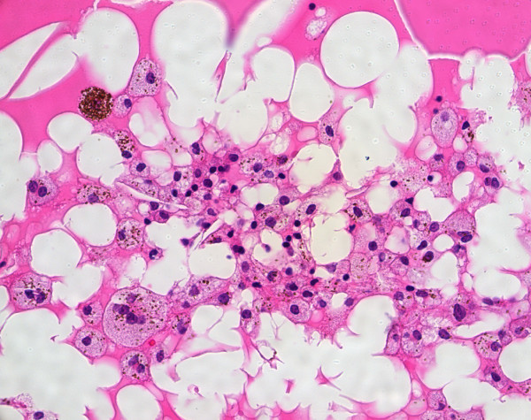
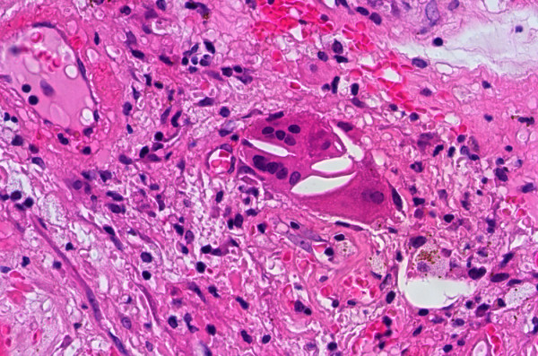
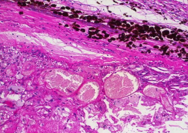
Case presentation: A 4 year-old girl presented with a blind and painful right eye. Ocular examination revealed neovascular glaucoma, cataract and posterior synechiae. Although viewing of the fundus was impossible, computed tomography disclosed total exudative retinal detachment in the affected eye. The eye was enucleated and subsequent histopathological evaluation confirmed the diagnosis of Coats' disease.
Conclusion: General pathologists usually do not have the opportunity to receive and study specimens from patients with Coats' disease. Coats' disease is one of the most important differential diagnoses of retinoblastoma. Therefore, it is crucial for the pathologist to be familiar with the histopathological features of the former, and distinguish it from the latter.
Figures
References
-
- Coats G. Forms of retinal diseases with massive exsudation. R Lond Ophthalmol Hosp Rep. 1908. pp. 440–525.
-
- Chang MM, McLean IW, Merritt JC. Coats' disease: a study of 62 histologically confirmed cases. J Pediatr Ophthalmol Strabismus. 1984;21:163–168. - PubMed
-
- Weller M, Bresgen M, Heimann K, Wiedemann P. [Immunohistology of proliferative vitreoretinopathy following giant tear detachment] Klin Monatsbl Augenheilkd. 1989;195:323–325. - PubMed
LinkOut - more resources
Full Text Sources
