. 2006 Nov;80(22):11355-61.
doi: 10.1128/JVI.00152-06. Epub 2006 Aug 30.

Rotavirus infection is not associated with small intestinal fluid secretion in the adult mouse

Affiliations

Rotavirus infection is not associated with small intestinal fluid secretion in the adult mouse

Shirin Kordasti et al. J Virol. 2006 Nov.

Abstract

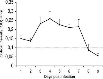
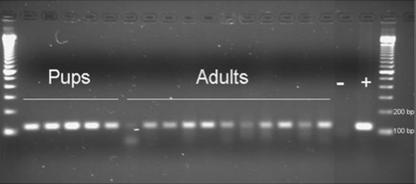
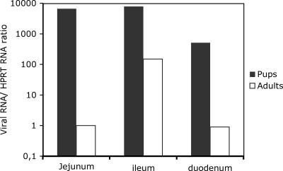
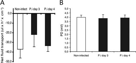
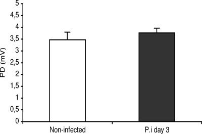
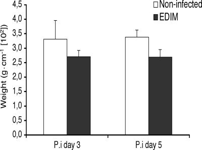
In contrast to humans, adult but not infant small animals are resistant to rotavirus diarrhea. The pathophysiological mechanism behind this age-restricted diarrhea is currently unresolved, and this question was investigated by studying the secretory state of the small intestines of adult mice infected with rotavirus. Immunohistochemistry and histological examinations revealed that rotavirus (strain EDIM) infects all parts of the small intestines of adult mice, with significant numbers of infected cells in the ilea at 2 and 4 days postinfection. Furthermore, quantitative PCR revealed that 100-fold more viral RNA was produced in the ilea than in the jejuna or duodena of adult mice. In vitro perfusion experiments of the small intestine did not reveal any significant changes in net fluid secretion among mice infected for 3 days or 4 days or in those that were noninfected (37 +/- 9 microl . h(-1) . cm(-1), 22 +/- 13 microl . h(-1) . cm(-1), and 33 +/- 6 microl . h(-1) . cm(-1), respectively) or in transmucosal potential difference (4.0 +/- 0.3 mV versus 3.9 +/- 0.4 mV), a marker for active chloride secretion, between control and rotavirus-infected mice. In vivo experiments also did not show any differences in potential difference between uninfected and infected small intestines. Furthermore, no significant differences in weight between infected and uninfected small intestines were found, nor were any differences in fecal output observed between infected and control mice. Altogether, these data suggest that rotavirus infection is not sufficient to stimulate chloride and water secretion from the small intestines of adult mice.


Figures

FIG. 1.
FIG. 1.
Shedding of rotavirus following infection with EDIM (n = 8). Values are means ± SEM. The horizontal line shows the optical density cutoff at 450 nm of 0.1.
FIG. 2.
FIG. 2.
Rotavirus PCR of stool samples collected 2 days p.i. from mouse pups (n = 5) and adult mice (n = 11) infected with EDIM rotavirus. Gene 6-specific primers were used to amplify a 118-bp PCR fragment. The amplicons were visualized with ethidium bromide on a 2% agarose gel. −, negative sample; +, positive sample.
FIG. 3.
FIG. 3.
Quantitative determination of rotavirus (EDIM) RNA in the intestines of infant and adult mice 4 days p.i., measured as the n-fold increase in viral RNA concentration compared to HPRT mRNA. The figure shows that the synthesis of viral RNA is higher in infant than in adult mice, relative to mRNA HPRT. The numbers presented are mean values (n = 3).
FIG. 4.
FIG. 4.
Immunohistochemistry of ileal tissue from infant and adult mice at 2 days p.i. Rotavirus-infected cells were identified with a rabbit polyclonal antiserum that recognizes predominantly vp6. (A) Immunoperoxidase staining of an infected ileum from an adult mouse. (B) A larger magnification of the tissue shown in panel A illustrating in more detail the infection of individual cells. (C) Immunoperoxidase staining of an ileal lesion from an adult mouse. Note the severe lesions proximate to the infected cells. (D) Immunoperoxidase staining of ileal tissue from an infected infant mouse with a large number of infected cells and severe vacuolization. Vacuoles in villous cells were a unique feature of infant mice.
FIG. 5.
FIG. 5.
In vitro perfusion experiments recording net fluid transport (A) and PD (B) in control segments and in infected segments at days 3 and 4 (n = 8 in each group). Values are means ± SEM.
FIG. 6.
FIG. 6.
In vivo recording of jejunal potential differences in control and rotavirus (EDIM)-infected mice at day 3 (n = 8 in each group). Values are means ± SEM.
FIG. 7.
FIG. 7.
Comparison of the small intestinal weights of control and rotavirus (EDIM)-infected mice at days 3 and 5 (n = 4 in each group). Values are means ± SEM.

References

    1. Anderson, E. J., and S. G. Weber. 2004. Rotavirus infection in adults. Lancet Infect. Dis. 4:91-99. - PMC - PubMed
    1. Banasaz, M., E. Norin, R. Holma, and T. Midtvedt. 2002. Increased enterocyte production in gnotobiotic rats mono-associated with Lactobacillus rhamnosus GG. Appl. Environ. Microbiol. 68:3031-3034. - PMC - PubMed
    1. Burns, J. W., A. A. Krishnaney, P. T. Vo, R. V. Rouse, L. J. Anderson, and H. B. Greenberg. 1995. Analyses of homologous rotavirus infection in the mouse model. Virology 207:143-153. - PubMed
    1. Ciarlet, M., M. E. Conner, M. J. Finegold, and M. K. Estes. 2002. Group A rotavirus infection and age-dependent diarrheal disease in rats: a new animal model to study the pathophysiology of rotavirus infection. J. Virol. 76:41-57. - PMC - PubMed
    1. Ciarlet, M., M. A. Gilger, C. Barone, M. McArthur, M. K. Estes, and M. E. Conner. 1998. Rotavirus disease, but not infection and development of intestinal histopathological lesions, is age restricted in rabbits. Virology 251:343-360. - PubMed

Publication types

MeSH terms