Comparative Study
. 2006 Nov;80(22):11000-8.
doi: 10.1128/JVI.01735-06. Epub 2006 Aug 30.

Crystal structure of west nile virus envelope glycoprotein reveals viral surface epitopes

Affiliations
Comparative Study

Crystal structure of west nile virus envelope glycoprotein reveals viral surface epitopes

Ryuta Kanai et al. J Virol. 2006 Nov.

Abstract

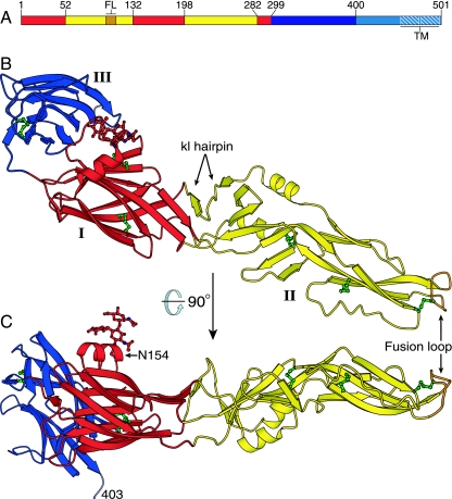
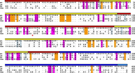
West Nile virus, a member of the Flavivirus genus, causes fever that can progress to life-threatening encephalitis. The major envelope glycoprotein, E, of these viruses mediates viral attachment and entry by membrane fusion. We have determined the crystal structure of a soluble fragment of West Nile virus E. The structure adopts the same overall fold as that of the E proteins from dengue and tick-borne encephalitis viruses. The conformation of domain II is different from that in other prefusion E structures, however, and resembles the conformation of domain II in postfusion E structures. The epitopes of neutralizing West Nile virus-specific antibodies map to a region of domain III that is exposed on the viral surface and has been implicated in receptor binding. In contrast, we show that certain recombinant therapeutic antibodies, which cross-neutralize West Nile and dengue viruses, bind a peptide from domain I that is exposed only during the membrane fusion transition. By revealing the details of the molecular landscape of the West Nile virus surface, our structure will assist the design of antiviral vaccines and therapeutics.


Figures

FIG. 1.
FIG. 1.
Structure of the West Nile virus sE monomer. (A) The three domains of West Nile virus E: domain I is red, domain II is yellow, and domain III is blue. The fusion loop (FL) is in orange. A 53-residue “stem” (cyan) links the ectodomain to a two-helix C-terminal transmembrane anchor (TM, white hatching). (B and C) The West Nile virus sE monomer, colored as in panel A, viewed in two perpendicular orientations. The last ordered residue in the sE structure (Ser403) and the kl hairpin, which forms the putative hydrophobic ligand-binding pocket, are labeled. The glycan at Asn154 and the six disulfide bonds are shown in ball-and-stick representation (in red and green, respectively).
FIG. 2.
FIG. 2.
Comparison of West Nile virus sE to other flavivirus sE structures. (A) Structure of dengue virus type 2 sE in the postfusion conformation (PDB entry 1OK8, in shades of gray) superimposed on West Nile virus sE (colored as in Fig. 1), using domain I as the reference. The C termini (in domain III) of the structures are 40 Å apart, while the fusion loops (in domain II) are less than 5 Å apart. The view is rotated 25° relative to panel B and Fig. 1B to show domain III of dengue virus sE more clearly. (B) Structure of the TBE virus sE dimer in the prefusion conformation (PDB entry 1SVB, in shades of gray) superimposed on West Nile virus sE, using domain I as the reference. Domains III of the structures are superimposed well, but the orientations of domain II are separated by a 20° rotation about a point near residue 196 (marked with a black and yellow star). This rotation translates into a 23-Å displacement of the fusion loop.
FIG. 3.
FIG. 3.
Structure-based alignment of the amino acid sequences of E proteins from West Nile virus (wn) strain 2741 (2), Japanese encephalitis virus (je) strain JaOArS982, dengue virus type 2 (d2) strain S1, and tick-borne encephalitis virus (tbe) strain Neudörfl. Dots indicate amino acid identities; dashes show gaps. The domains are indicated by a colored bar as in Fig. 1. The sequences are truncated at the last residue (406) of the soluble fragment (sE) of West Nile virus E, which we crystallized. The conserved glycosylation site in domain I is indicated by a red asterisk and red lettering. Residues lining the hydrophobic pocket in sE are shaded in gray. Residues that are exposed on the viral surface and are conserved in West Nile virus strains but not in other flaviviruses are shaded in magenta. Residues that are exposed on the viral surface and are conserved in wn, je, d2, and tbe viruses are shaded in orange.
FIG. 4.
FIG. 4.
Distribution of West Nile virus-specific residues on sE. (A and B) Two perpendicular views of West Nile virus sE, with residues that are conserved in West Nile virus strains but not in other flaviviruses shown in space-filling representation. Residues that are exposed on the surface of the mature virus are in magenta; residues that are not exposed are in gray. The I0-strand peptide recognized by recombinant antibodies scFv-Fc 11, 71, and 73 (residues 281 to 300) is shown in green, with the two essential basic residues (Lys287 and Lys291) in space-filling representation. Most West Nile virus-specific neutralizing antibodies bind an epitope that includes Thr330 (21, 31, 36). The view in panel B is perpendicular to the viral surface, such that the outside of the virion is up. The views are the same as in Fig. 1B and C. (C) Atomic model of the West Nile virus outer protein shell based on the 9.5-Å-resolution electron cryomicroscopic reconstruction of dengue virus (41). E assembles into dimers in mature virions. The glycan of West Nile virus E is shown in red, residues lining the putative hydrophobic pocket in dark gray, residues 281 to 300 (a partial epitope of scFv-Fcs 11, 71, and 73) in green, and the epitope of therapeutic antibody E16 (32) in blue. The fusion loop is in orange. A black triangle connects the icosahedral symmetry axes. (D) Close-up of panel C, with the two-, three-, and fivefold icosahedral symmetry axes labeled. A single sE monomer is circled with a semitransparent gray line. The minimum separation between glycans (red) is ∼50 Å.
FIG. 5.
FIG. 5.
Partial epitope mapping of therapeutic recombinant antibodies previously selected by phage display (13). To map linear peptide sequences of scFv-Fcs 11, 71, 73, 79, and 95, we measured scFv-Fc binding to a set of overlapping 20-mer peptides spanning the entire sequence of West Nile virus sE by ELISA. scFv-Fcs 11, 71, and 73 bind a peptide consisting of West Nile virus residues 281 to 300. All three scFv-Fcs also recognize the homologous E peptide from dengue virus type 2 (DEN2) but not a peptide composed of the same amino acids in a randomized sequence. Furthermore, scFv-Fc binding was critically dependent on positively charged side chains (Lys or Arg) at positions 287 and 291 of West Nile virus E.

References

    1. Allison, S. L., J. Schalich, K. Stiasny, C. W. Mandl, C. Kunz, and F. X. Heinz. 1995. Oligomeric rearrangement of tick-borne encephalitis virus envelope proteins induced by an acidic pH. J. Virol. 69:695-700. - PMC - PubMed
    1. Anderson, J. F., T. G. Andreadis, C. R. Vossbrinck, S. Tirrell, E. M. Wakem, R. A. French, A. E. Garmendia, and H. J. Van Kruiningen. 1999. Isolation of West Nile virus from mosquitoes, crows, and a Cooper's hawk in Connecticut. Science 286:2331-2333. - PubMed
    1. Beasley, D. W., M. C. Whiteman, S. Zhang, C. Y. Huang, B. S. Schneider, D. R. Smith, G. D. Gromowski, S. Higgs, R. M. Kinney, and A. D. Barrett. 2005. Envelope protein glycosylation status influences mouse neuroinvasion phenotype of genetic lineage 1 West Nile virus strains. J. Virol. 79:8339-8347. - PMC - PubMed
    1. Bressanelli, S., K. Stiasny, S. L. Allison, E. A. Stura, S. Duquerroy, J. Lescar, F. X. Heinz, and F. A. Rey. 2004. Structure of a flavivirus envelope glycoprotein in its low-pH-induced membrane fusion conformation. EMBO J. 23:728-738. - PMC - PubMed
    1. Brünger, A. T., P. D. Adams, G. M. Clore, W. L. DeLano, P. Gros, R. W. Grosse-Kunstleve, J. S. Jiang, J. Kuszewski, M. Nilges, N. S. Pannu, R. J. Read, L. M. Rice, T. Simonson, and G. L. Warren. 1998. Crystallography & NMR system: a new software suite for macromolecular structure determination. Acta Crystallogr. D 54:905-921. - PubMed

Publication types

Associated data

LinkOut - more resources