. 2006 Oct;17(10):2706-18.
doi: 10.1681/ASN.2006040412. Epub 2006 Aug 30.

Polycystin-2 immunolocalization and function in zebrafish

Affiliations

Polycystin-2 immunolocalization and function in zebrafish

Tomoko Obara et al. J Am Soc Nephrol. 2006 Oct.

Abstract

Polycystin-2 functions as a cation-permeable transient receptor potential ion channel in kidney epithelial cells and when mutated results in human autosomal dominant polycystic kidney disease. For further exploration of the in vivo functions of Polycystin-2, this study examined its expression and function during zebrafish embryogenesis. pkd2 mRNA is ubiquitously expressed, and its presence in the larval kidney could be confirmed by reverse transcription-PCR on isolated pronephroi. Immunostaining with anti-zebrafish Polycystin-2 antibody revealed protein expression in motile kidney epithelial cell cilia and intracellular cell membranes. Intracellular localization was segment specific; in the proximal nephron segment, Polycystin-2 was localized to basolateral cell membranes, whereas in the caudal pronephric segment, Polycystin-2 was concentrated in subapical cytoplasmic vesicles. Polycystin-2 also was expressed in muscle cells and in a variety of sensory cells that are associated with mechanotransduction, including cells of the ear, the lateral line organ, and the olfactory placodes. Disruption of Polycystin-2 mRNA expression resulted in pronephric kidney cysts, body axis curvature, organ laterality defects, and hydrocephalus-defects that could be rescued by expression of a human PKD2 mRNA. In-frame deletions in the first extracellular loop and C-terminal phosphofurin acidic cluster sorting protein-1 (PACS-1) binding sites in the cytoplasmic tail caused Polycystin-2 mislocalization to the apical cell surface. Unlike zebrafish intraflagellar transport protein (IFT) mutants, cyst formation was not associated with cilia defects and instead correlated with reduced kidney fluid output, expansion of caudal duct apical cell membranes, and occlusion of the caudal pronephric nephron segment.


Figures
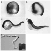
Figure 1
Figure 1
Expression of pkd2 mRNA during embryogenesis. By whole-mount in situ hybridization, pkd2 is expressed ubiquitously during epiboly (A), at the 18-somite stage (B), at 24 h postfertilization (hpf; C) and at 48 hpf (D). Reverse transcription–PCR (RT-PCR) on RNA from isolated pronephric ducts (E) confirmed expression of pkd2 in pronephroi.
Figure 2
Figure 2
Immunolocalization of Polycystin-2 in the pronephric kidney. (A) Expression of Polycystin-2 protein in the trunk of a 48-hpf embryo viewed in whole mount is strong in muscle (*) and the pronephric duct (arrowheads). (B) Immunizing peptide preincubation control for antibody staining shows no signal in a similar trunk region (*muscle; arrowheads, pronephric duct). (C) Control for antibody specificity using ATG morpholino (MO) blockade of endogenous protein translation shows no expression in the trunk region (*muscle; arrowheads, pronephric duct). (D) Z-series projection of Polycystin-2 immunofluorescence in anterior pronephric duct shows expression in cilia and associated with basolateral cell membranes and infoldings (arrows) in a whole mount–stained 2.5-d embryo. (E) Cross-sections of the anterior pronephric duct show Polycystin-2 expression associated with basolateral cell membranes (arrows) and apical cilia. (F) Polycystin-2 immunofluorescence in the posterior pronephric duct is punctate and distributed throughout the cells with a concentration of staining near the apical surface. (G) Sections of the posterior pronephric duct in the cloaca region show expression of Polycystin-2 in subapical membrane vesicles and reduced expression in basolateral membranes (arrows). (H) Western blot of adult zebrafish kidney reacted with anti-zebrafish PKD2 antibody reveals a single band migrating at approximately 115 kD.
Figure 3
Figure 3
Nephron segment-specific distribution of Polycystin-2 in basolateral membranes and cilia. (A through C) The anterior pronephric duct. Polycystin-2 (A; green) and acetylated tubulin (B; red) immunofluorescence in confocal sections. (C) Merged image of A and B. Anterior duct Polycystin-2 is present associated with basolateral membranes (arrows in A) and a lumenal bundle of cilia. (D through F) The posterior pronephric duct. Polycystin-2 (D; green) and acetylated tubulin (E; red) immunofluorescence in confocal sections. (F) Merge of D and E shows accumulation of Polycystin-2 in intracellular vesicles and at the base of cilia (arrows). (G through I) The anterior pronephric duct. (G) Tangential confocal section through the basolateral membranes shows a concentration of punctate Polycystin-2 expression in a whole mount-stained 2.5-d embryo. (H) Basolateral membranes of the anterior duct stain uniformly with the monoclonal α6F against the NaK ATPase α1 subunit. (I) Merge of G and H shows that Polycystin-2 and the NaK ATPase show areas of co-localization and some distinct areas of punctate expression. (J through L) The posterior pronephric duct. (J) Cross-section of the duct shows punctate Polycystin-2 immunofluorescence in lateral and apical membranes but absence of expression in basal cell surfaces. A lumenal cilium also is positive for Polycystin-2. (K) NaK ATPase immunofluorescence on basolateral membranes. (L) Merge of J and K.
Figure 4
Figure 4
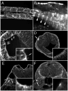
Immunolocalization of Polycystin-2 in nonkidney organs. (A) At 56 hpf, muscle cell membranes of the trunk myotomes are positive for Polycystin-2 in confocal sections. (B) Polycystin-2 expression also is present in repeating, sarcomeric bands on intracellular muscle fibers. (C) Ependymal cell cilia in the brain ventricles are positive for Polycystin-2 (arrows and inset) in histologic sections. (D) The olfactory placode cell membranes are strongly positive for Polycystin-2 (arrow) in histologic sections. (Inset) Confocal section of the olfactory placode showing concentration of Polycystin-2 immunoreactivity in apical membranes of olfactory placode cells. (E) Epithelial cells of the ear show apical staining for Polycystin-2 (Inset) Higher magnification view of ear cells showing Polycystin-2 immunoreactivity in apical membranes and vesicles. (F) Cells of the lateral line organ are strongly positive for Polycystin-2 expression (Inset) Higher magnification view of lateral line organ cells in the central part of this structure showing Polycystin-2 expression.
Figure 5
Figure 5
Disruption of pkd2 expression with splice donor antisense MO oligonucleotides. (A) Diagram of Polycystin-2 protein in the cell membrane showing membrane topology and C-terminal motifs associated with Polycystin-2 function. (B) Targeting splice donor sites of exons 3, 5, 12, and 13 with antisense MO oligonucleotides resulted in the production of internal in-frame deletions (exons 3, 5, and 13 MO) and a C-terminal truncation (exon 12 MO). Transmembrane domains of Polycystin-2 are depicted in gray. (C) The efficacy of the injected MO was quantified at 24-, 48-, and 72-h intervals by RT-PCR. Polycystin-2 exon 3 donor MO (MOex3) caused a 39–amino acid in-frame deletion of part of the first transmembrane domain as detected by the presence of a smaller, internally deleted RT-PCR product. Some recovery of normal Polycystin-2 mRNA was observed by 72 hpf. Similar results were observed for all splice donor targeted MO. (D) Control embryos at 72 hpf. (E) Histologic section of normal pronephros at 72 hpf. (F) MOex3-injected embryo showing axis curvature and hydrocephalus (arrow; 97%, 941 of 967 embryos). (G) Histologic section showing cystic pronephric tubules (*) in MOex3-injected embryo. (H) MOex12-injected embryos and section (I) of the cystic pronephros. (J) MOex13-injected embryos showing severe axis curvature and kidney cysts (arrow). (K) Histologic section of embryo in J showing kidney cyst (*). (L) Antisense MO deletions in the Polycystin-2 protein sequence predicted on the basis of nucleotide sequence of RT-PCR products amplified from MO-injected embryos. In-frame deletions are shown in gray for MOex3, MOex5, and MOex13. MOex12 induced a nonsplicing event that resulted in a stop codon immediately after exon 12 in the cDNA (shown in red *). Transmembrane domains (tm) and the PKD1 binding homology domain are highlighted in brown. Membrane targeting motifs in the cytoplasmic C-terminus including the phosphofurin acidic cluster sorting protein–1 (PACS-1)-binding acidic cluster are underlined.
Figure 6
Figure 6
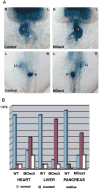
Polycystin-2 function in left–right asymmetry. (A) Expression of cardiac myosin light chain 2 (cmlc2) in control embryos (top left) demonstrates normal positions of the heart ventricle (v) and atrium (a) relative to the embryo midline. Polycystin-2 MOex3 caused inversion of left–right axes in 50% of injected embryos (top left). Forkhead2 (fkd2) and insulin gene expression revealed similar inversion of liver (li) and pancreas (p) situs, respectively (bottom). (B) Quantification of left–right asymmetry defects. Wild-type embryos (wt) show normal situs in most all cases with a low level of background laterality defects. Polycystin-2 MOex3-injected embryos show a high degree of left–right axis inversion (approximately 50%) as well as midline organ position for the heart, liver, and pancreas.
Figure 7
Figure 7
Rescue of pkd2MOex3 phenotype with human PKD2 mRNA co-injection. (A and E) pkd2 MOex3-injected embryo showing axis curvature and pronephric cysts in cross-section (97%; 617 of 638 injected embryos). (B and F) Co-injection of 30 pg of human PKD2 mRNA with MOex3 MO results in partial rescue of cyst phenotype (arrow in F; 98%, 571 of 583 injected embryos). (C and G) Co-injection with 100 pg of human PKD2 mRNA completely rescues cyst phenotype (arrow in G; 99%, 728 of 733 embryos). (D and H) Injection of human PKD2 mRNA alone has no effect on normal embryos (641 embryos). (I) Polyclonal anti-PKD2 antibody 96526 recognizes mouse and human Polycystin-2 but not zebrafish Polycystin-2 (uninjected wild-type embryo). (J) Embryo injected with human PKD2 mRNA shows broad expression of the exogenous rescuing mRNA. (K) Mouse Polycystin-2 peptide antigen preincubation blocks all 96526 antibody staining, demonstrating specific immunoreactivity of the 96526 antibody.
Figure 8
Figure 8
Apical mislocalization of MO-altered Polycystin-2 proteins. (A) Wild-type Polycystin-2 is concentrated in basolateral membranes of the anterior pronephric ducts (arrows, basolateral cell surface) in histologic sections. (B) MOex5-injected embryo showing a shift in immunoreactivity of altered (in-frame deletion in the first extracellular loop) Polycystin-2 from basal to apical cell membranes. (C) MOex13-injected embryo showing absence of basolateral staining and concentration of Polycystin-2 immunoreactivity in apical and lumenal membranes.
Figure 9
Figure 9
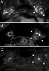
Reduction in pronephric fluid output in pkd2 morphants. (A and B) Pronephric fluid output detection at the cloaca using rhodamine dextran as a fluid tracer in wild-type embryos at 72 hpf. Appearance of a fluorescence “jet” of urine output at the cloaca is observed (arrowhead in B). (C and D) Polycystin-2 MOex3-injected embryos at 72 hpf show a significant reduction in fluid output at the cloaca (arrowhead in D).
Figure 10
Figure 10
Structural alterations in the distal pronephric nephron segment of pkd2 morphants. (A) Differential interference contrast images of the cloaca region of the pronephric duct in living, 3-dpf wild-type embryos show a patent lumen extending to the cloaca. (B) Polycystin-2 MOex3-injected embryos show an apparent collapse of the distal duct lumen. (C) Lumenal distension (*) immediately anterior to an area of duct occlusion (arrowhead in C) in a Polycystin-2 MOex3-injected embryo. Cilia beat and length appeared normal in areas of lumenal distension (data not shown). (H) Electron micrograph cross-section of the wild-type distal pronephric duct showing a patent duct lumen (*). For reference, cell basement membrane is denoted with arrowheads. (I) A similar region of the pronephric duct in a Polycystin-2 MOex3-injected embryos appears occluded by extended apical cytoplasm of duct epithelial cells. Five apical adherens junctions of the MOex3 duct epithelial cells can be seen (white arrowheads); however, the apical membranes of these cells abut with opposing cells, occluding the duct lumen. Black arrowheads, basement membrane.

Similar articles

Cited by

References

    1. Clapham DE. TRP channels as cellular sensors. Nature. 2003;426:517–524. - PubMed
    1. Cai Y, Maeda Y, Cedzich A, Torres VE, Wu G, Hayashi T, Mochizuki T, Park JH, Witzgall R, Somlo S. Identification and characterization of polycystin-2, the PKD2 gene product. J Biol Chem. 1999;274:28557–28565. - PubMed
    1. Foggensteiner L, Bevan AP, Thomas R, Coleman N, Boulter C, Bradley J, Ibraghimov-Beskrovnaya O, Klinger K, Sandford R. Cellular and subcellular distribution of polycystin-2, the protein product of the PKD2 gene. J Am Soc Nephrol. 2000;11:814–827. - PubMed
    1. Koulen P, Cai Y, Geng L, Maeda Y, Nishimura S, Witzgall R, Ehrlich BE, Somlo S. Polycystin-2 is an intracellular calcium release channel. Nat Cell Biol. 2002;4:191–197. - PubMed
    1. Nauli SM, Alenghat FJ, Luo Y, Williams E, Vassilev P, Li X, Elia AE, Lu W, Brown EM, Quinn SJ, Ingber DE, Zhou J. Polycystins 1 and 2 mediate mechanosensation in the primary cilium of kidney cells. Nat Genet. 2003;33:129–137. - PubMed

Publication types

Substances