. 2006 Sep;26(18):6936-49.
doi: 10.1128/MCB.01040-06.

Evidence for the requirement of ITAM domains but not SLP-76/Gads interaction for integrin signaling in hematopoietic cells

Affiliations

Evidence for the requirement of ITAM domains but not SLP-76/Gads interaction for integrin signaling in hematopoietic cells

Farhad Abtahian et al. Mol Cell Biol. 2006 Sep.

Abstract

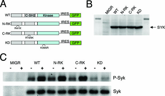
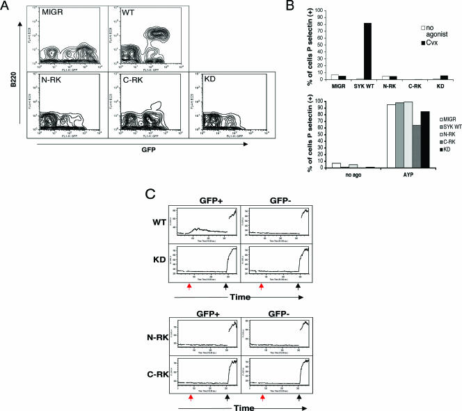
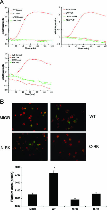
Syk tyrosine kinase and Src homology 2 (SH2) domain-containing leukocyte-specific phosphoprotein of 76 kDa (SLP-76) are signaling mediators activated downstream of immunoreceptor tyrosine-based activation motif (ITAM)-containing immunoreceptors and integrins. While the signaling cascades descending from integrins are similar to immunoreceptors, the mechanism of Syk activation and SLP-76 recruitment remains unclear. We used an in vivo structure-function approach to study the requirements for the domains of Syk and SLP-76 in immunoreceptor and integrin signaling. We found that both SH2 domains and the kinase domain of Syk are required for immunoreceptor-dependent signaling and cellular response via integrins. While the Gads-binding domain of SLP-76 is needed for immunoreceptor signaling, it appears dispensable for integrin signaling. Syk and SLP-76 also are required for initiating and/or maintaining separation between the blood and lymphatic vasculature. Therefore, we correlated the signaling requirement of the various domains of Syk and SLP-76 to their requirement in regulating vascular separation. Our data suggest ITAMs are required in Syk-dependent integrin signaling, demonstrate the separation of the structural features of SLP-76 to selectively support immunoreceptor versus integrin signaling, and provide evidence that the essential domains of SLP-76 for ITAM signals are those which most efficiently support separation between lymphatic and blood vessels.


Figures

FIG. 1.
FIG. 1.
SH2 domains of Syk are dispensable for activation by αIIbβ3 in CHO cells. (A) Schematic of Syk constructs. N-RK and C-RK contain arginine-to-lysine mutations at amino acids 41 and 194, respectively, and KD contains a lysine-to-arginine mutation at amino acid 395. (B) CHO cells were transiently transfected with empty vector, WT Syk, or Syk mutant. Syk expression was assayed by Western blotting after normalizing for the percentage of CHO cells transfected. (C) CHO cells were transiently transfected with the MIGR vector expressing either Syk or a Syk mutant. Cells were plated upon fibrinogen-coated (+) or BSA-coated (−) plates, and lysates from equivalent numbers of transfected cells were collected and immunoprecipitated for Syk, followed by Western blotting for phosphotyrosine. Blots were then stripped and reprobed for Syk.
FIG. 2.
FIG. 2.
Mutation in either SH2 domain of Syk results in loss of ITAM-dependent signaling in vivo. FLCs from Syk-deficient embryos were transduced with virus expressing GFP alone (MIGR1) or GFP with WT Syk or mutant Syk and were used to reconstitute lethally irradiated C57BL6 mice. Eight to 10 weeks postreconstitution, chimeric mice were sacrificed for study. (A) Splenocytes from reconstituted mice were isolated and stained for B220, followed by flow cytometric analysis to distinguish GFP-positive from GFP-negative cells. Note that B cells (upper quadrants) appear only when WT Syk is successfully transduced. (B) Retrovirally transduced platelets were either left unstimulated or stimulated with 5 ng CVX (top) or 0.5 mM AYPGKF (AYP) peptide (bottom). GFP-positive platelets were then assessed for P-selectin expression. Results are plotted as the percentage of platelets staining positive for P-selectin. (C) Neutrophils were labeled with Indo-1 and incubated with an antibody against FcRγ. Calcium flux was measured by flow cytometry following cross-linking of the receptors with anti-rat immunoglobulin G (red arrow) and ionomycin (black arrow).
FIG. 3.
FIG. 3.
Mutation in either SH2 domain of Syk results in loss of the integrin-dependent cellular response in vivo. (A) Neutrophils from chimeric mice were stimulated with TNF or left unstimulated after plating upon a fibrinogen-coated surface. ROI production was measured by cytochrome c oxidation. Each graph includes neutrophils expressing WT Syk (red line) and one Syk mutant. (B) Platelets were incubated on coverslips coated with fibrinogen, fixed, permeabilized, and stained for F-actin (phalloidin). GFP-positive platelets represent platelets transduced with virus. Original magnification, ×1,000. The average area of spread of GFP+ platelets (n > 100) is shown in pixels per platelet. Error bars represent standard errors. *, significant difference in spreading between MIGR and WT, N-RK, or C-RK (P < 0.001).
FIG. 4.
FIG. 4.
Expression of WT SLP-76, mutant SLP-76, and MTS. (A) Schematic of WT and mutant GFP-tagged SLP and raft-targeted SLP-76 (MTS). PRD represents the proline-rich domain of SLP-76. For the Y3F mutant, stars represent mutations of tyrosine to phenylalanine at amino acids 112, 128, and 145. For the G2 mutant, stars represent amino acid changes from arginine to alanine at amino acid 237 and lysine to alanine at position 240. MTS consists of the first 35 amino acids of LAT fused to full-length SLP-76. MTS is not GFP tagged and hence runs at a lower molecular weight. (B) CHO cells were transiently transfected with empty vector or vector expressing WT SLP-76, mutant SLP-76, or MTS. Cell lysates of equivalent numbers of transfected cells were examined by immunoblot analysis with anti-SLP-76 antibody.
FIG. 5.
FIG. 5.
Membrane localization of SLP-76 is required for immunoreceptor-dependent signaling but is dispensable for an integrin-dependent response. FLCs or bone marrow cells from SLP-76-deficient embryos were transduced with virus expressing empty vector or WT SLP-76, mutant SLP-76, or MTS and were used to reconstitute lethally irradiated C57BL6 mice. Eight to 10 weeks postreconstitution, chimeric mice were sacrificed for study. (A) Retrovirally transduced platelets were either left unstimulated or stimulated with 5 nM CVX (top) or 0.5 mM AYPGKF (AYP) (bottom) for 20 min in the presence of anti-P-selectin antibody. P-selectin expression was assessed by flow cytometry with gates set to distinguish GFP-positive cells from GFP-negative cells. Results are plotted as the percentage of GFP-positive platelets staining positive for P-selectin. (B) Neutrophils from chimeric mice were stimulated with TNF or left unstimulated after plating upon a fibrinogen-coated surface. ROI production was measured by cytochrome c oxidation. (C) Platelets were incubated on coverslips coated with fibrinogen, fixed, permeabilized, and stained for F-actin (phalloidin). Original magnification, ×1,000. The average area of spread of GFP+ platelets (n > 100) is shown in pixels per platelet. Error bars represent standard errors. formula image
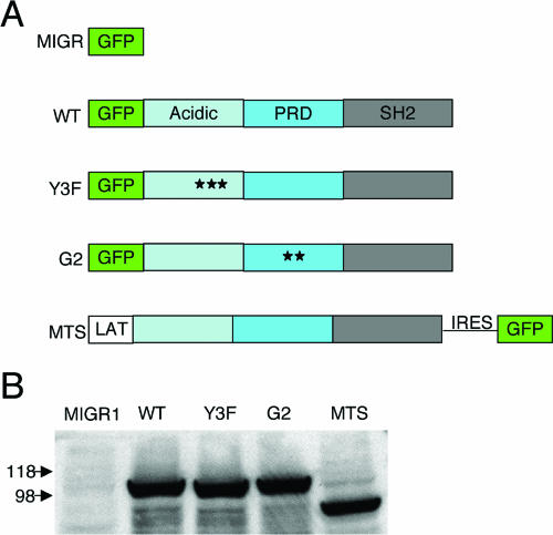
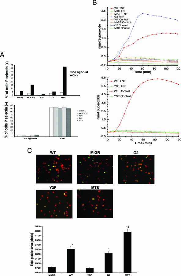
, significant difference in spreading between MIGR or Y3F and WT or G2 (P < 0.001); #, significant difference in spreading between MTS and WT (P < 0.001).
FIG. 6.
FIG. 6.
SH2 domains and kinase activity of Syk are required for blood-lymphatic separation. (A) Range of the gross morphological appearances of vascular phenotypes seen in the small intestines of chimeric mice: −, unaffected; +, blood visible in mesenteric lymph nodes; ++, focal loss of normal vascular architecture of gut wall; +++, complete loss of vascular architecture with appearance of chylous effusion within peritoneum. (B) Chimeric mice were sacrificed and examined for appearance of grossly visible vascular lesions in the small intestine and mesenteric lymph nodes. Black arrows identify affected regions. (C) Tissue from the small intestine of chimeric mice was fixed, paraffin embedded, sectioned, and stained for Lyve-1 with horseradish peroxidase-tagged secondary antibody to identify lymphatic vessels. Arrows identify lymphatic vessels. Original magnification: (A and B) ×1, except for C-RK which was at ×2; (C) ×200.
FIG. 7.
FIG. 7.
Membrane localization of SLP-76 is required for blood-lymphatic vascular separation. (A) Chimeric mice were sacrificed and examined for the appearance of grossly visible vascular lesions in the small intestine and mesenteric lymph nodes. Original magnification, ×1. (B) Tissue from small intestines of chimeric mice was fixed, paraffin embedded, sectioned, and stained for Lyve-1 with horseradish peroxidase-tagged secondary antibody to identify lymphatic vessels. Arrows identify lymphatic vessels. Original magnification, ×200.

References

    1. Abtahian, F., A. Guerriero, E. Sebzda, M. M. Lu, R. Zhou, A. Mocsai, E. E. Myers, B. Huang, D. G. Jackson, V. A. Ferrari, V. Tybulewicz, C. A. Lowell, J. J. Lepore, G. A. Koretzky, and M. L. Kahn. 2003. Regulation of blood and lymphatic vascular separation by signaling proteins SLP-76 and Syk. Science 299:247-251. - PMC - PubMed
    1. Asada, H., N. Ishii, Y. Sasaki, K. Endo, H. Kasai, N. Tanaka, T. Takeshita, S. Tsuchiya, T. Konno, and K. Sugamura. 1999. Grf40, a novel Grb2 family member, is involved in T cell signaling through interaction with SLP-76 and LAT. J. Exp. Med. 189:1383-1390. - PMC - PubMed
    1. Banerji, S., J. Ni, S. X. Wang, S. Clasper, J. Su, R. Tammi, M. Jones, and D. G. Jackson. 1999. LYVE-1, a new homologue of the CD44 glycoprotein, is a lymph-specific receptor for hyaluronan. J. Cell Biol. 144:789-801. - PMC - PubMed
    1. Boerth, N. J., J. J. Sadler, D. E. Bauer, J. L. Clements, S. M. Gheith, and G. A. Koretzky. 2000. Recruitment of SLP-76 to the membrane and glycolipid-enriched membrane microdomains replaces the requirement for linker for activation of T cells in T cell receptor signaling. J. Exp. Med. 192:1047-1058. - PMC - PubMed
    1. Bourette, R. P., S. Arnaud, G. M. Myles, J. P. Blanchet, L. R. Rohrschneider, and G. Mouchiroud. 1998. Mona, a novel hematopoietic-specific adaptor interacting with the macrophage colony-stimulating factor receptor, is implicated in monocyte/macrophage development. EMBO J. 17:7273-7281. - PMC - PubMed

Publication types

MeSH terms

LinkOut - more resources