. 2006 Sep;26(18):6993-7004.
doi: 10.1128/MCB.01060-06.

Functional requirement of noncoding Y RNAs for human chromosomal DNA replication

Affiliations

Functional requirement of noncoding Y RNAs for human chromosomal DNA replication

Christo P Christov et al. Mol Cell Biol. 2006 Sep.

Abstract

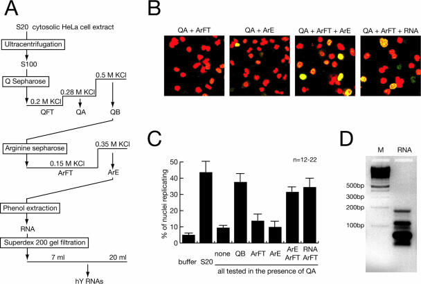
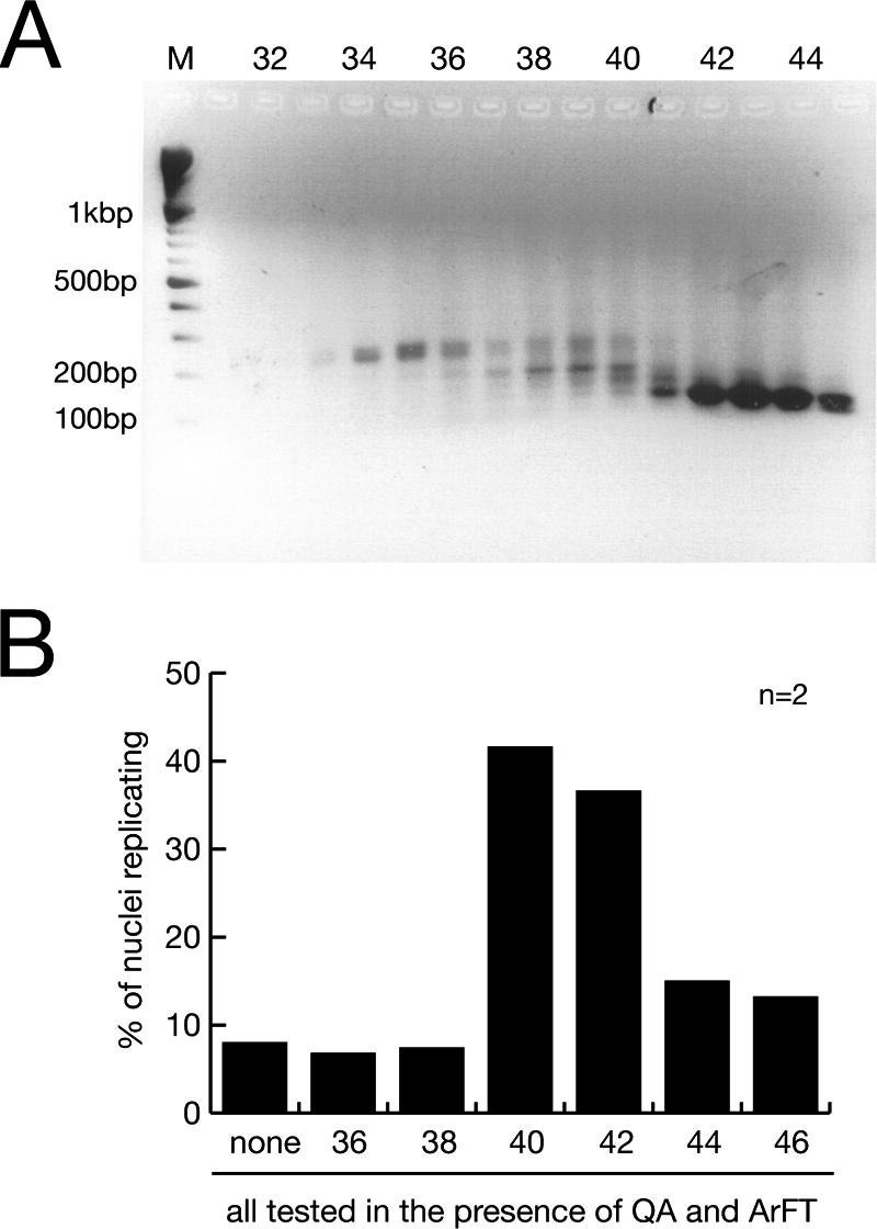
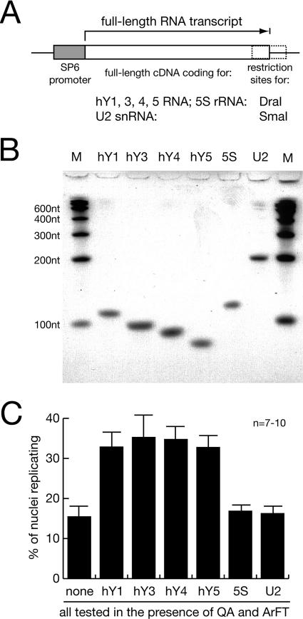
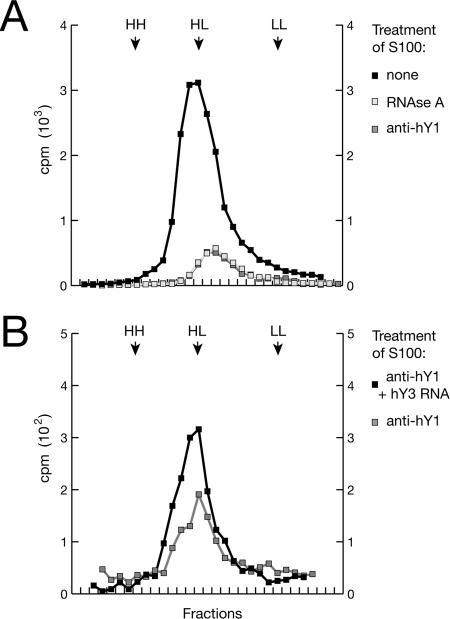
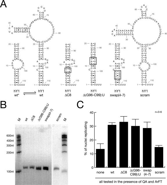
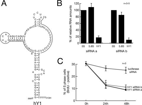
Noncoding RNAs are recognized increasingly as important regulators of fundamental biological processes, such as gene expression and development, in eukaryotes. We report here the identification and functional characterization of the small noncoding human Y RNAs (hY RNAs) as novel factors for chromosomal DNA replication in a human cell-free system. In addition to protein fractions, hY RNAs are essential for the establishment of active chromosomal DNA replication forks in template nuclei isolated from late-G(1)-phase human cells. Specific degradation of hY RNAs leads to the inhibition of semiconservative DNA replication in late-G(1)-phase template nuclei. This inhibition is negated by resupplementation of hY RNAs. All four hY RNAs (hY1, hY3, hY4, and hY5) can functionally substitute for each other in this system. Mutagenesis of hY1 RNA showed that the binding site for Ro60 protein, which is required for Ro RNP assembly, is not essential for DNA replication. Degradation of hY1 RNA in asynchronously proliferating HeLa cells by RNA interference reduced the percentages of cells incorporating bromodeoxyuridine in vivo. These experiments implicate a functional role for hY RNAs in human chromosomal DNA replication.


Figures

FIG. 1.
FIG. 1.
Purification of RNA as a factor for human chromosomal DNA replication. (A) Schematic diagram of fractionation steps. (B) Representative fields of nuclei replicating in vitro. Template nuclei from late-G1-phase cells were incubated with combinations of the indicated fractions, and replicating nuclei were detected by confocal fluorescence microscopy as detailed in references and . Nuclear DNA is visualized by propidium iodide (red signal). Replicated DNA is labeled by incorporation of digoxigenin-dUMP, which is detected by fluorescein-conjugated antidigoxigenin Fab fragments (green signal). A merged signal appears in yellow. (C) Quantitative analysis of replicating G1-phase nuclei in vitro. Mean values and standard deviations of the proportions of replicating nuclei from the indicated reactions of 12 to 22 independent experiments (n) are shown (see Materials and Methods). Protein amounts per experiment were 100 μg unfractionated S20 cytosolic extract, 15 μg QA, 35 μg QB, 8 μg ArFT, a 20-μl volume of concentrated ArE containing less than 0.1 μg protein or the equivalent RNA prepared from this volume as specified. Fractions were used as indicated. (D) Visualization of RNA purified from fraction ArE. The RNA present in fraction ArE was purified by phenol extraction and isopropanol precipitation, separated on a 2% neutral agarose gel, and visualized by staining with ethidium bromide (lane RNA). A ladder of multimeric 100-bp DNA fragments was used as a molecular weight marker (lane M). An inverted image of the fluorescent gel is shown.
FIG. 2.
FIG. 2.
A discrete class of RNAs is essential for human chromosomal DNA replication. (A) Size fractionation of RNA prepared from ArE by gel filtration. Individual fractions were analyzed by agarose gel electrophoresis, and a 100-bp DNA ladder was used as a molecular weight marker (lane M). (B) Functional testing of the size-fractionated RNA. n, number of experiments. Template nuclei were incubated with the RNA purified from a constant volume of the indicated gel filtration fractions, supplemented with fractions QA and ArFT. Proportions of replicating nuclei were determined as described in the legend to Fig. 1.
FIG. 3.
FIG. 3.
Nucleotide (nt) sequences and predicted secondary structures of human Y RNAs (28, 41). Nucleotide sequences complementary to antisense DNA oligonucleotides (see below; see also Fig. 5) are highlighted by gray lines.
FIG. 4.
FIG. 4.
Human Y RNA is required for the reconstitution of chromosomal DNA replication. (A) Schematic drawing of the expression constructs for full-length human RNAs. (B) In vitro synthesis of full-length human wild-type RNA. Individual recombinant RNAs were synthesized in vitro from the constructs shown in panel A and visualized by SYBR green staining after denaturing gel electrophoresis. Multimeric 100-nucleotide (nt) RNA fragments were used for a molecular weight marker (lane M). (C) Functional reconstitution of chromosomal DNA replication with hY RNAs. n, number of experiments. Nuclei from late-G1-phase cells were incubated with fractions QA and ArFT, supplemented with 100 ng of the individual RNAs synthesized in vitro as indicated. Proportions of replicating nuclei were determined as described in the legend to Fig. 1.
FIG. 5.
FIG. 5.
Degradation of hY RNAs in vitro inhibits chromosomal DNA replication in late-G1-phase template nuclei. n, number of experiments. (A) Specific degradation of hY RNA in vitro. S100 cytosolic extract from proliferating HeLa cells was treated with RNase A or with the DNA antisense oligonucleotides complementary to single-stranded domains of the hY RNAs as shown in Fig. 3. As a negative control, the standard bacteriophage T3 DNA sequencing primer was used. The proportions of the indicated relative RNA amounts remaining in the extract after the treatment were determined by quantitative RT-PCR, using 5S rRNA as the reference. (B) Y RNA degradation reduces the proportion of nuclei replicating in vitro. Late-G1-phase template nuclei were incubated in untreated and treated S100 cytosolic extract as indicated. Proportions of replicating nuclei were determined as described in the legend to Fig. 1. (C) Y RNA degradation reduces the amount of extract-dependent DNA synthesis in late-G1-phase nuclei in vitro. Nuclei were incubated in untreated and treated S100 cytosolic extract in the presence of [α-32P]dCTP as indicated. The incorporation of dNMPs into nascent chromosomal DNA under these conditions was quantitated by precipitation with trichloroacetic acid and scintillation counting. (D) Functional substitution of one depleted hY RNA in the extract by a different hY RNA. After depletion of either hY1 or hY3 RNA by antisense DNA oligonucleotides, the treated extract was supplemented with 100 ng of the indicated hY RNAs synthesized in vitro. Proportions of replicating nuclei were determined as described in the legend to Fig. 1.
FIG. 6.
FIG. 6.
Human Y RNAs are required for semiconservative DNA replication. Late-G1-phase nuclei were incubated with S100 cytosolic extract in the presence of BrdU triphosphate and [α-32P]dCTP for 3 h, and purified DNA reaction products were analyzed by density gradient centrifugation and scintillation counting. (A) Specific degradation of hY RNA inhibits semiconservative DNA replication. Cytosolic extract from proliferating HeLa cells was left untreated, treated with RNase A, or treated with anti-hY1 oligonucleotides. Positions of unsubstituted (light-light [LL]), hemisubstituted (heavy-light [HL]), and fully substituted DNA (heavy-heavy [HH]) are indicated as determined by refractive indices. (B) Reconstitution of semiconservative DNA replication by hY RNA. After depletion of hY1 RNA by anti-hY1 oligonucleotides, the treated extract was supplemented with 100 ng of hY3 RNA synthesized in vitro. Representative results of one out of two independent experiments are shown (note that quantitative comparisons of the raw cpm values should not be drawn between the independent experiments of panels A and B).
FIG. 7.
FIG. 7.
The conserved binding site for Ro60 protein on hY1 RNA is not essential for chromosomal DNA replication. scram, scrambled nucleotide sequence mutant. (A) Nucleotide sequences and predicted structures of wild-type (hY1wt) and mutant hY1 RNAs. Nucleotide reference numbers for the RNAs are indicated by gray numbers. Structure predictions were performed using the RNAfold algorithm (http://rna.tbi.univie.ac.at) (12). Two alternative and interconvertible structural configurations of the wt stem are shown on the left (hY1wt and hY1wt*). The stems of three mutants deficient in binding to Ro60 protein (10) are shown in the middle [hY1ΔC8, hY1Δ(G96-C99):U, and hY1swap(4-7)]. The mutated binding sites for Ro60 are boxed. A scrambled nucleotide sequence mutant that maintains the overall predicted structure is shown on the left (hY1scram). The connecting wt loop is not drawn for some mutants. (B) In vitro synthesis of full-length mutant hY1 RNAs. Individual recombinant RNAs are visualized after denaturing gel electrophoresis as detailed in the legend to Fig. 3B. nt, nucleotides. (C) Functional reconstitution of chromosomal DNA replication with mutant hY1 RNAs. n, number of experiments. Nuclei from late-G1-phase cells were incubated in fractions QA and ArFT, supplemented with 100 ng of the individual mutant hY1 RNAs as indicated. Proportions of replicating nuclei were determined as described in the legend to Fig. 1C.
FIG. 8.
FIG. 8.
Degradation of hY1 RNA in human cells by RNA interference in vivo leads to an inhibition of DNA replication. n, number of experiments. (A) Generation of siRNAs specific for hY1 RNA. The nucleotide sequences complementary to the two siRNAs (termed a and b) are indicated by gray lines. (B) Quantification of RNA levels after RNAi against hY1 RNA. At 48 h after transfection of asynchronously proliferating HeLa cells with the indicated siRNAs, the expression levels of hY1 RNA and 5.8S rRNA, relative to 5S rRNA, were determined by quantitative real-time RT-PCR. Relative RNA amounts were normalized against transfection with a nontarget siRNA specific for firefly luciferase mRNA. (C) Quantification of replicating S-phase cells after RNAi. At the indicated times after transfection with the indicated siRNAs, cells were pulse labeled for 1 h with BrdU, and percentages of BrdU-positive cells were determined by immunofluorescence microscopy. Mean values and standard deviations of three to five independent experiments are shown as indicated.

References

    1. Arias, E. E., and J. C. Walter. 2004. Initiation of DNA replication in xenopus egg extracts. Front. Biosci. 9:3029-3045. - PubMed
    1. Bernstein, E., and C. D. Allis. 2005. RNA meets chromatin. Genes Dev. 19:1635-1655. - PubMed
    1. Chen, X., A. M. Quinn, and S. L. Wolin. 2000. Ro ribonucleoproteins contribute to the resistance of Deinococcus radiodurans to ultraviolet irradiation. Genes Dev. 14:777-782. - PMC - PubMed
    1. Chen, X., and S. L. Wolin. 2004. The Ro 60 kDa autoantigen: insights into cellular function and role in autoimmunity. J. Mol. Med. 82:232-239. - PubMed
    1. Coverley, D., H. Laman, and R. A. Laskey. 2002. Distinct roles for cyclins E and A during DNA replication complex assembly and activation. Nat. Cell Biol. 4:523-528. - PubMed

Publication types

MeSH terms