. 2006 Aug 30;26(35):9006-9.
doi: 10.1523/JNEUROSCI.2370-06.2006.

Altered hippocampal synaptic potentiation in P2X4 knock-out mice

Affiliations

Altered hippocampal synaptic potentiation in P2X4 knock-out mice

Joan A Sim et al. J Neurosci. .

Abstract

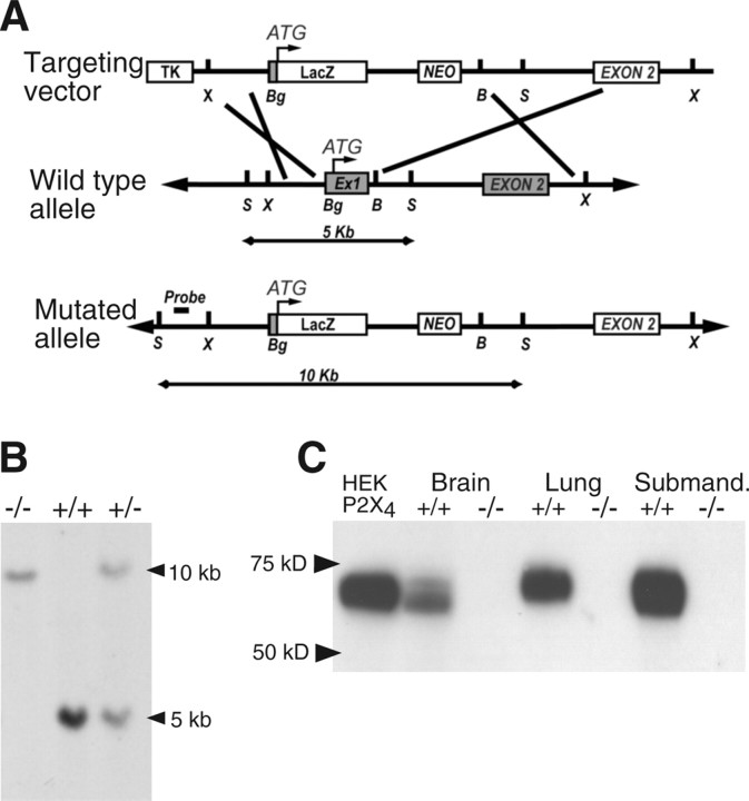
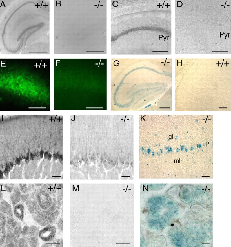
P2X4 purinergic receptors are calcium-permeable, ATP-activated ion channels. In the CA1 area of the hippocampus, they are located at the subsynaptic membrane somewhat peripherally to AMPA receptors. The possible role of P2X4 receptors has been difficult to elucidate because of the lack of selective antagonists. Here we report the generation of a P2X4 receptor knock-out mouse and show that long-term potentiation (LTP) at Schaffer collateral synapses is reduced relative to that in wild-type mice. Ivermectin, which selectively potentiates currents at P2X4, was found to increase LTP in wild-type mice but had no effect in P2X4 knock-out mice. We suggest that calcium entry through subsynaptic P2X4 receptors during high-frequency stimulation contributes to synaptic strengthening.


Figures

Figure 1.
Figure 1.
Targeted disruption of the mouse P2X4 gene. A, Maps of the P2X4 wild-type allele, targeting vector, and mutant allele. Boxes represent open reading frames; LacZ gene insertion upstream of the P2X4-initiating methionine resulted in the deletion of all the coding region of exon 1 and of the first exon–intron splice site. TK, Thymidine kinase; NEO, neomycin; Ex1, exon 1; B, BamH1; Bg, BgIII; S, SacI; X, XbaI. Southern blotting probe was designed outside of the targeting locus between 5′ SacI and XbaI sites. As shown, after digestion with SacI, this probe will hybridize to a 5 or 10 kb DNA fragment for wild-type and mutant allele, respectively. B, Southern blot analysis of genomic DNA digested with SacI from +/+, +/−, and −/− mice using the probe described in A. C, Western blot of membrane protein fraction from brain, lung, and submandibular gland of wild-type P2X4+/+ mice showed a band at 60 kDa, similar to that observed in P2X4-expressing HEK293 cells. No band of equivalent size was observed from knock-out P2X4−/− mice.
Figure 2.
Figure 2.
Expression of P2X4 receptors in wild-type P2X4+/+ but not in knock-out P2X4−/− mice. A, C, E, P2X4 subunit immunoreactivity throughout cell body layers of hippocampus is absent in knock-out mice (B, D, F). Pyr, Pyramidal cell. G, H, Detection of β-galactosidase. 5-Bromo-4-chloro-3-indolyl-β-d-galactopyranoside (X-gal) staining in hippocampus of knock-out P2X4−/− mice but not in wild-type P2X4+/+ mice. Scale bars: A, B, G, H, 500 μm; C, D, 50 μm; E, F, 40 μm. I, P2X4 subunit immunoreactivity in cerebellar Purkinje cells is absent in the knock-out P2X4−/− mice (J). K, X-gal staining shows the expression of β-galactosidase in cerebellar Purkinje cells (P), as well as some cells of the granular layer (gl) and mossy layer (ml) from the knock-out P2X4−/− mouse. L, P2X4 subunit immunoreactivity in acinar and ductal cells of the submandibular gland is completely absent in the knock-out P2X4−/− mouse (M). N, Extensive expression of β-galactosidase in submandibular gland of the knock-out P2X4−/− mouse. Scale bars: I, J, K, 20 μm; L, M, N, 50 μm.
Figure 3.
Figure 3.
Contribution of P2X4 receptor activation to tetanus-induced LTP in hippocampal CA1 pyramidal cells. A, The magnitude of LTP elicited by Schaffer collateral stimulation was significantly reduced in P2X4−/− mice (▵; n = 12) compared with wild-type P2X4+/+ mice (○; n = 12; p < 0.001). B, LTP is enhanced by ivermectin (3 μm; ·) in wild-type P2X4+/+ mice but not in knock-out P2X4−/− mice (C) (▴; n = 15; p > 0.05), because they were superimposable on control values (▵). Ivermectin was applied 10 min before tetanic stimulation (downward filled arrows); it did not change the baseline of field EPSP. Tetanic stimulation (4 times at 100 Hz for 1 s) was applied at the upward open arrows. All data points are mean ± SEM, for n mice. Control data points in A are the same as in B and C.

References

    1. Anderson WW, Collingridge GL. The LTP Program: a data acquisition program for on-line analysis of long-term potentiation and other synaptic events. J Neurosci Methods. 2001;108:71–83. - PubMed
    1. Bliss TVP, Collingridge GL. A synaptic model of memory: long-term potentiation in the hippocampus. Nature. 1993;361:31–39. - PubMed
    1. Buell G, Lewis C, Collo G, North RA, Surprenant A. An antagonist-insensitive P2X receptor expressed in epithelia and brain. EMBO J. 1996;15:55–62. - PMC - PubMed
    1. Collo G, North RA, Kawashima E, Merlo-Pich E, Neidhart S, Surprenant A, Buell G. Cloning of P2X5 and P2X6 receptors and the distribution and properties of an extended family of ATP-gated ion channels. J Neurosci. 1996;16:2495–2507. - PMC - PubMed
    1. Conquet F. Inactivation in vivo of metabotropic glutamate receptor 1 by specific chromosomal insertion of reporter gene lacZ. Neuropharmacology. 1995;34:865–870. - PubMed

Publication types

MeSH terms