. 2006 Aug 30;26(35):9047-56.
doi: 10.1523/JNEUROSCI.2797-06.2006.

Glucocorticoids increase amyloid-beta and tau pathology in a mouse model of Alzheimer's disease

Affiliations

Glucocorticoids increase amyloid-beta and tau pathology in a mouse model of Alzheimer's disease

Kim N Green et al. J Neurosci. .

Abstract

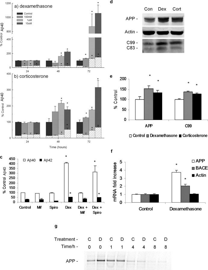
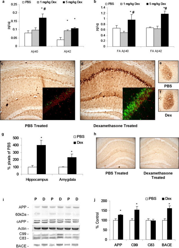
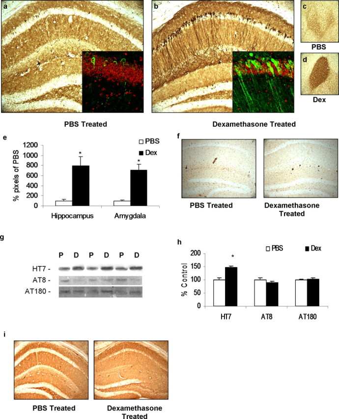
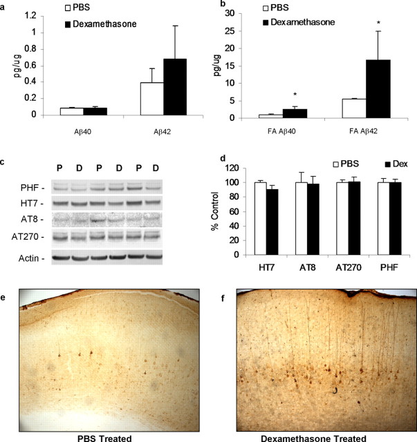
Various environmental and genetic factors influence the onset and progression of Alzheimer's disease (AD). Dysregulation of the hypothalamic-pituitary-adrenal (HPA) axis, which controls circulating levels of glucocorticoid hormones, occurs early in AD, resulting in increased cortisol levels. Disturbances of the HPA axis have been associated with memory impairments and may contribute to the cognitive decline that occurs in AD, although it is unknown whether such effects involve modulation of the amyloid beta-peptide (Abeta) and tau. Using in vitro and in vivo experiments, we report that stress-level glucocorticoid administration increases Abeta formation by increasing steady-state levels of amyloid precursor protein (APP) and beta-APP cleaving enzyme. Additionally, glucocorticoids augment tau accumulation, indicating that this hormone also accelerates the development of neurofibrillary tangles. These findings suggest that high levels of glucocorticoids, found in AD, are not merely a consequence of the disease process but rather play a central role in the development and progression of AD.


Figures

Figure 1.
Figure 1.
Glucocorticoids enhance Aβ production in a time- and concentration-dependent manner. N2A cells were incubated with dexamethasone (100 nm to 10 μm; n = 3 per condition) (a) or corticosterone (100 nm to 10 μm; n = 3 per condition) (b) for 24–72 h, and Aβ levels were measured from cell media by sandwich ELISA. Aβ42 levels were expressed as a percentage of control Aβ40, shown in the thatched area. *p < 0.05, significance versus controls for either Aβ40 or Aβ42. Control Aβ40 levels average out at ∼1 pg of Aβ40 microgram of protein for a 24 h period into 1 ml of media. c, N2A cells were incubated for 72 h with or without mifepristone (Mif; 10 μm; n = 3) or spironolactone (Spiro; 100 nm; n = 3) and in the presence or absence of 1 μm dexamethasone (Dex; n = 3 per condition), and media Aβ levels were measured. *p < 0.05, significance versus controls for either Aβ40 or Aβ42. d, Treatment of N2A cells with dexamethasone (Dex; 1 μm, 48 h; n = 3) or corticosterone (Cort; 5 μm, 48 h; n = 3) or control (Con; 48 h; n = 3) increases steady-state levels of full-length APP and selectively increases in C99 as shown by Western blot. e, Quantification of d with protein levels normalized to β-actin levels as a loading control. *p < 0.05, significance versus controls. f, mRNA levels of mouse APP and BACE are increased after 72 h treatment with 10 μm dexamethasone, as measured by real-time PCR. *p < 0.05, significance versus controls. g, Pulse chase analyses of 35S-labeled APP after treatment of N2A cells with 10 μm dexamethasone for 72 h (D; n = 3) or control (C; n = 3). Cells were pulsed with 35S, after starvation, for 1 h and chased at the 0, 1, 4, and 8 h time points.
Figure 2.
Figure 2.
Dexamethasone treatment increases Aβ, C99, and BACE in the 3×Tg-AD mouse model. Four-month-old male 3×Tg-AD mice were treated daily for 7 d with 1 mg/kg (n = 5) or 5 mg/kg (n = 8) dexamethasone (Dex) or PBS vehicle (n = 8). a, Detergent-soluble Aβ levels were measured in whole-brain homogenates. Mice treated with 5 mg/kg dexamethasone (7 d; black bars) had significantly higher levels of Aβ40 and Aβ42 than vehicle-treated (PBS, 7 d; white bars) mice, whereas mice treated with 1 mg/kg dexamethasone (7 d; gray bars) had elevated Aβ42 compared with PBS controls. *p < 0.05, significance versus PBS controls for either Aβ40 or Aβ42; #p < 0.05, significance versus 1 mg/kg dexamethasone treatment. b, Detergent-insoluble Aβ40 and Aβ42 levels were significantly increased between 5 mg/kg dexamethasone treated and vehicle treated. c, e, DAB staining with 6E10 shows Aβ-like immunoreactivity in 40 μm sections from vehicle-treated mice. Hippocampus and amygdala regions are shown at 10× magnification. Inset demonstrates 40× confocal images with Aβ-like immunoreactivity shown in green and nuclear stain TOTO red shown in red. d, f, Same staining but in dexamethasone-treated animals. Aβ-like immunoreactivity was elevated in cell bodies of the hippocampus, as also shown by confocal imaging. g, Quantification of 6E10 DAB immunoreactivity in hippocampal and amygdala regions from PBS-treated and 5 mg/kg dexamethasone-treated groups. h, DAB staining with anti-CD45 shows that no activated microglia could be detected in the hippocampus of either PBS- or dexamethasone-treated animals. i, Western blot analyses of protein extracts from whole-brain homogenates of dexamethasone- and vehicle-treated 3×Tg-AD mice, shown as alternating lanes: P, PBS vehicle; D, 5 mg/kg dexamethasone treated. j, Quantification of protein blots from i shown normalized to β-actin levels as a loading control. Steady-state levels of APP are increased in the dexamethasone-treated group (6E10), whereas a 60 kDa band is similarly decreased, suggesting alternative processing. cAPP levels are unaltered, whereas C99 levels are increased but C83 levels remain unchanged. Furthermore, steady-state levels of BACE protein are increased concomitant to C99 levels. *p < 0.05, significance versus PBS controls.
Figure 3.
Figure 3.
Dexamethasone (Dex) treatment in 3×Tg-AD mice increases tau accumulation. Four-month-old male 3×Tg-AD mice were treated daily for 7 d with 5 mg/kg dexamethasone (n = 8) or PBS vehicle (n = 8). a, c, Immunostaining with anti-tau antibody HT7 shows little immunoreactivity in 40 μm sections from PBS vehicle-treated mice. Hippocampus and amygdala regions are shown at 10× magnification. Inset demonstrates 40× confocal images with HT7 immunoreactivity shown in green and nuclear stain TOTO red shown in red. b, d, Same staining but in 5 mg/kg dexamethasone-treated animals. HT7 immunoreactivity was elevated in cell bodies of the hippocampus and throughout the neuronal processes, as also shown by confocal imaging. e, Quantification of HT7 DAB immunoreactivity in hippocampal and amygdala regions from PBS-treated and 5 mg/kg dexamethasone-treated groups. f, DAB staining of no primary controls shown from hippocampus of PBS-treated and 5 mg/kg dexamethasone-treated animals, 10×. g, Western blot analyses of protein extracts from whole-brain homogenates of dexamethasone- and vehicle-treated 3×Tg-AD mice, shown as alternating lanes (P, PBS vehicle; D, 5 mg/kg dexamethasone treated) showing increases in total human tau steady-state levels (HT7) but no differences in phospho-tau (AT8 and AT180). h, Quantification of protein blots from g shown normalized to β-actin levels as a loading control. i, 2×Tg mice (PS1M146VKI and tauP301L, lacking the human APP transgene) treated daily with dexamethasone (5 mg/kg, 7 d; n = 3) or vehicle (PBS, 7 d; n = 3). Staining with HT7 shows no immunoreactivity in 40 μm sections from vehicle-treated or 5 mg/kg dexamethasone-treated mice. Hippocampus region is shown at 10× magnification. DAB HT7 immunoreactivity was unchanged from vehicle.
Figure 4.
Figure 4.
Dexamethasone (Dex) treatment increases insoluble Aβ in 13-month-old 3×Tg-AD mice. Thirteen-month-old male 3×Tg-AD mice were treated daily for 7 d with 5 mg/kg dexamethasone (n = 4) or PBS vehicle (n = 4). a, Detergent-soluble Aβ levels were measured in whole-brain homogenates. Mice treated with dexamethasone (5 mg/kg, 7 d; black bars) had significantly higher levels of Aβ40 and Aβ42 than vehicle treated (PBS, 7 d; white bars). b, Detergent-insoluble Aβ levels were significantly different between dexamethasone treated and vehicle treated. *p < 0.05, significance versus PBS controls for either Aβ40 or Aβ42. c, Western blot analyses of protein extracts from whole-brain homogenates of 13-month-old dexamethasone- and vehicle-treated 3×Tg-AD mice, shown as alternating lanes (P, PBS vehicle; D, 5 mg/kg dexamethasone treated) showing no differences in total or phosphorylated-tau (PHF). d, Quantification of protein blots from c shown normalized to β-actin levels as a loading control. e, Immunostaining with anti-tau antibody HT7 shows little immunoreactivity in 40 μm sections from 13-month-old PBS vehicle-treated 3×Tg-AD mice. Cortical region is shown at 10× magnification. f, Same staining but in dexamethasone-treated animals. DAB HT7 immunoreactivity was notably elevated in cell bodies and processes in the cortex, which was absent in PBS-treated vehicles.
Figure 5.
Figure 5.
Plasma corticosterone levels from 3×Tg-AD mice compared with NonTg over time. a, Plasma obtained from 3×Tg-AD and NonTg mice was taken and corticosterone levels were measured at 2, 4, 6, 9, 12, 15, and 18 months of age. No significant changes are seen at 2, 4, and 6 months. At 9 months and older, 3×Tg-AD mice have significantly higher plasma corticosterone levels than NonTg mice. *p < 0.05, significance versus NonTg animals at that time point. b, Western blot analyses of protein extracts from hippocampal homogenates of 4- and 15-month-old NonTg and 3×Tg-AD mice (n = 4 per group), shown as alternating lanes (C, NonTg control; 3, 3×Tg-AD), showing no differences in glucocorticoid receptor (GR) steady-state levels. c, Quantification of protein blots from b shown normalized to β-actin levels as a loading control.

References

    1. Abraham I, Harkany T, Horvath KM, Veenema AH, Penke B, Nyakas C, Luiten PG. Chronic corticosterone administration dose-dependently modulates Abeta(1–42)- and NMDA-induced neurodegeneration in rat magnocellular nucleus basalis. J Neuroendocrinol. 2000;12:486–494. - PubMed
    1. Aisen PS, Davis KL, Berg JD, Schafer K, Campbell K, Thomas RG, Weiner MF, Farlow MR, Sano M, Grundman M, Thal LJ. A randomized controlled trial of prednisone in Alzheimer’s disease. Alzheimer’s Disease Cooperative Study. Neurology. 2000;54:588–593. - PubMed
    1. Billings LM, Oddo S, Green KN, McGaugh JL, Laferla FM. Intraneuronal Abeta causes the onset of early Alzheimer’s disease-related cognitive deficits in transgenic mice. Neuron. 2005;45:675–688. - PubMed
    1. Braak H, Braak E. Diagnostic criteria for neuropathologic assessment of Alzheimer’s disease. Neurobiol Aging. 1997;18:S85–S88. - PubMed
    1. Budas G, Coughlan CM, Seckl JR, Breen KC. The effect of corticosteroids on amyloid beta precursor protein/amyloid precursor-like protein expression and processing in vivo. Neurosci Lett. 1999;276:61–64. - PubMed

Publication types

MeSH terms