. 2006 Aug 23:12:983-94.

Targeted inhibition of p57 and p15 blocks transforming growth factor beta-inhibited proliferation of primary cultured human limbal epithelial cells

Affiliations

Targeted inhibition of p57 and p15 blocks transforming growth factor beta-inhibited proliferation of primary cultured human limbal epithelial cells

Zhuo Chen et al. Mol Vis. .

Abstract

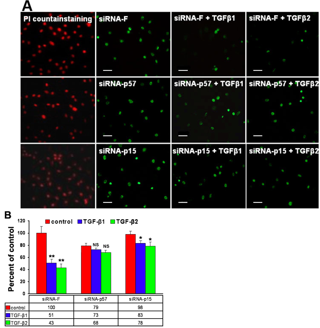
Purpose: To evaluate the role of cyclin-dependent kinase inhibitors p57 and p15 in transforming growth factor (TGF)-beta1 or TGF-beta2 inhibited proliferation of primary cultured human limbal epithelial cells using short interfering RNA (siRNA).

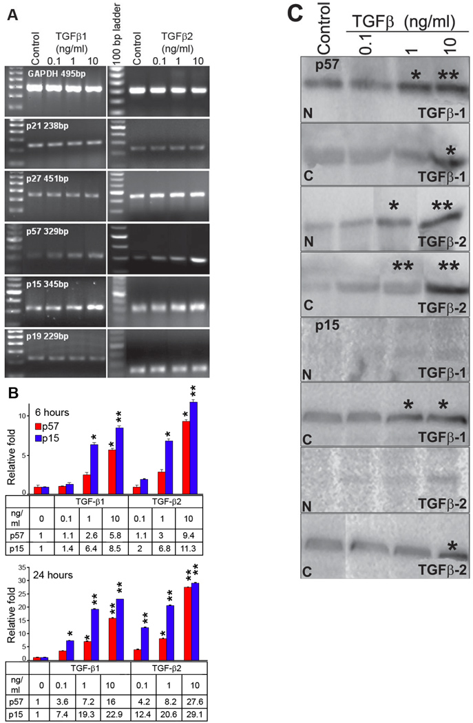
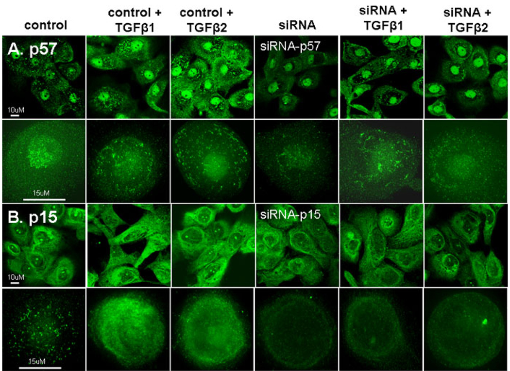
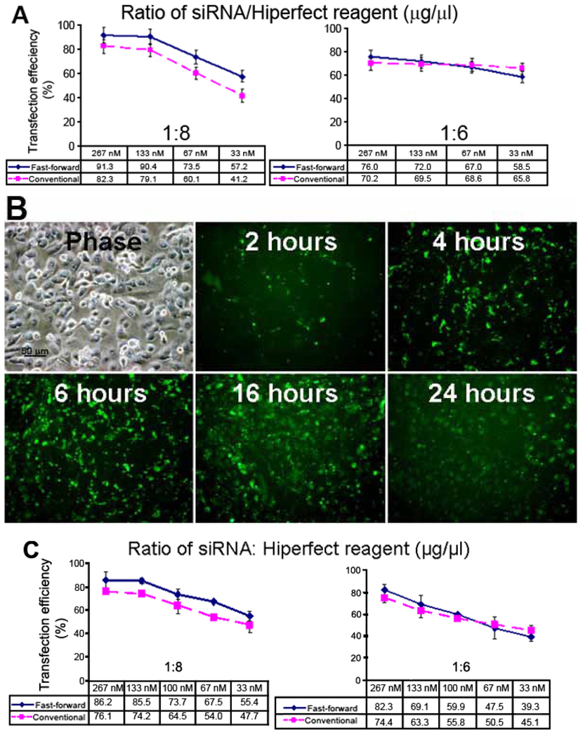
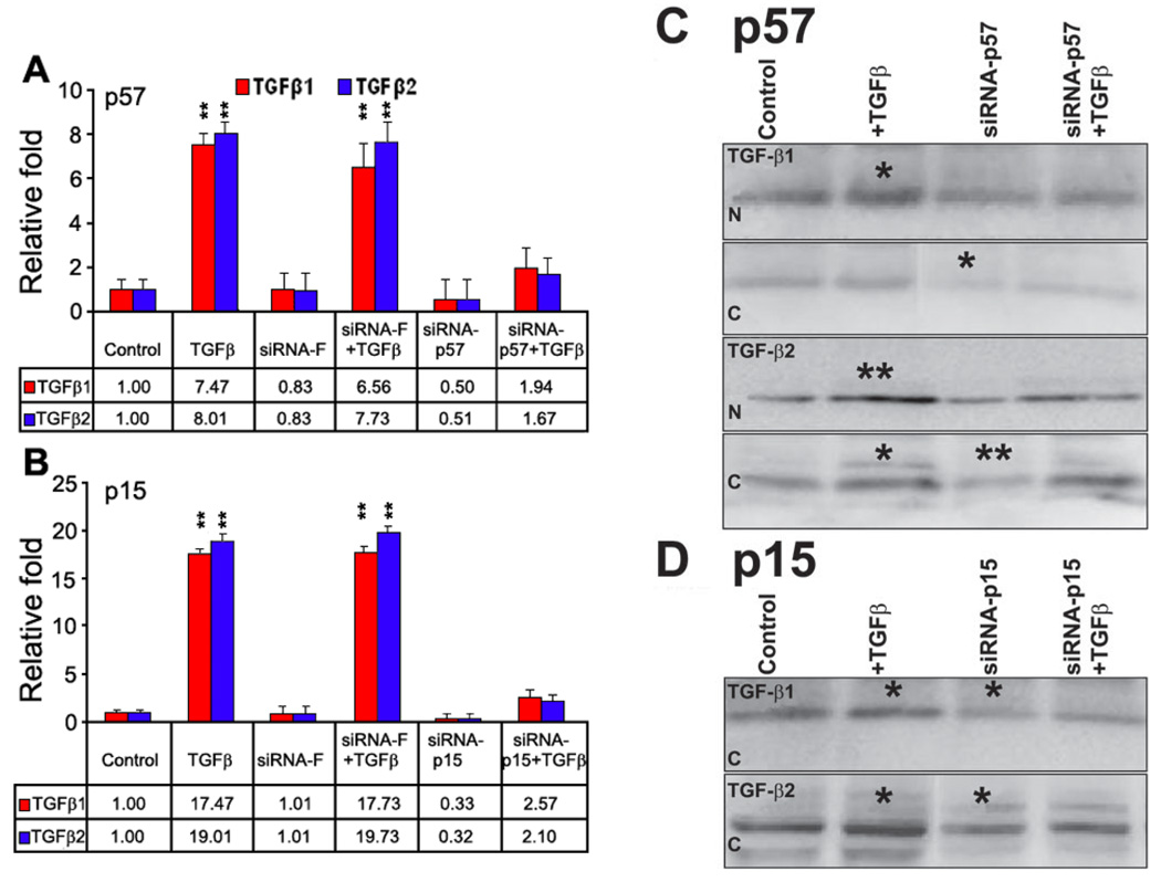
Methods: Primary cultured human limbal epithelial cells were treated with TGF-beta1 or TGF-beta2 for 6 and 24 h, and total RNA extracted for RT-PCR and real-time PCR using primers for p21, p27, and p57 (CipP/Kip family) and p15 and p19 (INK4 family). Proteins were extracted for western blot analysis of p57 and p15. For RNA interference, primary cultured human limbal epithelial cells were transfected with annealed double-stranded siRNA (67 nM) specific for p57, p15, or siRNA-Fluorescein (siRNA-F; as a negative control) followed by treatment with TGF-beta1 or TGF-beta2 at 1 ng/ml. P57 and p15 were quantitatively detected by real-time PCR and western blot; and immunolocalized by immunofluorescent staining. The effects of TGF-beta1 or TGF-beta2 on cell proliferation were evaluated by BrdU incorporation and MTT assay.

Results: TGF-beta1 or TGF-beta2 significantly inhibited primary cultured human limbal epithelial cell proliferation measured by BrdU incorporation and MTT assay. TGF-beta1 or TGF-beta2 upregulated the expression of p57 and p15 mRNA and protein, but did not effect the expression of p19, p21, or p27. The siRNA transfection efficiency of these cells was 75% and no cellular toxicity was observed by 24 h. The TGF-beta1 or TGF-beta2 stimulated expression of p57 and p15 mRNA were markedly blocked by siRNA-p57 or siRNA-p15, respectively, but not by siRNA-F. The TGF-beta1 or TGF-beta2 suppression of epithelial proliferation measured by BrdU incorporation and MTT generation was increased to near normal levels by siRNA-p57 or siRNA-p15. Western blot and immunofluorescent staining showed that levels of p57 and p15 proteins were equally reduced in the cytoplasm and nucleus.

Conclusions: These findings demonstrate that TGF-beta1 and/or TGF-beta2 inhibit proliferation of primary cultured human limbal epithelial cells and that p57 and p15 play roles in this process.


Figures

Figure 1
Figure 1
TGF-β1 and TGF-β2 regulation of cyclin-dependent kinase inhibitors (CDKI) in primary cultured human limbal epithelial cells. A: Semi-quantitative RT-PCR of different doses of TGF-β1 and TGF-β2 treatment for 6 h. B: Relative quantitative real-time PCR for p57 and p15 in primary cultured human limbal epithelial cells before and following TGF-β1 and TGF-β2 treatment for 6 and 24 h. The asterisk indicates a p<0.05 and the double asterisk indicates a p<0.01. C: Western blot analysis showed the location of p57 and p15 before and following TGF-β1 and TGF-β2 treatment of nuclear (N) and cytoplasmic (C) fractions for 24 h. The asterisk indicates a p<0.05 and the double asterisk indicates a p<0.01.
Figure 2
Figure 2
Immunofluorescent staining, LSCM (first row) and deconvolution microscopy (second row) for A: p57 in control, siRNA-p57 treated before and following TGF-β1 or TGF-β2 treated (1 ng/ml) in primary cultured human limbal epithelial cells. B: p15 in control, siRNA-p57 treated before and following TGF-β1 or TGF-β2 treated in primary cultured human limbal epithelial cells.
Figure 3
Figure 3
Transfection efficiency of different ratios of siRNA/Hiperfect reagent and at different times. A: Comparison of transfection efficiency of siRNA-F in primary cultured human limbal epithelial cells using Fast-Forward and conventional methods with different concentrations of siRNA-F and different ratios of siRNA to Hiperfect reagent by flow cytometry. B: Fluorescent microscopy showing fluorescent (siRNA-F) transfection efficiency at different times following the Fast-Forward technique using 67 nM with a 1:8 ratio of siRNA to transfection reagent (µg/µl). C: Comparison of transfection efficiency of siCONTROL TOX in primary cultured human limbal epithelial cells using Fast-Forward and conventional methods with different concentrations of siCONTROL TOX and different ratios of siCONTROL TOX to Hiperfect reagent.
Figure 4
Figure 4
mRNA and protein analysis of primary cultured human limbal epithelial cells followed by TGF-β treatment after siRNA-p15 and siRNA-p57. A: Relative quantitative real-time PCR analysis showing the effects of siRNA-F and siRNA-p57 on p57 mRNA expression in control and TGF-β (1 ng/ml) stimulated primary cultured human limbal epithelial cells. B: Relative quantitative real-time PCR analysis showing the effects of siRNA-F and siRNA-p15 on p15 mRNA expression in control and TGF-β stimulated (1 ng/ml) primary cultured human limbal epithelial cells. C: Western blot analysis of p57 protein in nuclear (N) and cytoplasmic (C) preparations following TGF-β1 and TGF-β2 treatment (1 ng/ml) for 24 h in primary cultured human limbal epithelial cells with or without siRNA-p57. D: Western blot analysis of p15 protein in cytoplasmic preparation following TGF-β1 and TGF-β2 treatment (1 ng/ml) for 24 h in primary cultured human limbal epithelial cells with or without siRNA-p15. No p15 protein was detected in the nucleus, as shown in Figure 1C. In both C and D, the asterisk indicates a p<0.05 and the double asterisk indicates a p<0.01.
Figure 5
Figure 5
Cell proliferation ability assay. A: Representative BrdU incorporation in primary cultured human limbal epithelial cells treated with siRNA-F, siRNA-p57, and siRNA-p15 with or without TGF-β1 or TGF-β2 treatment (1 ng/ml). B: MTT assay showing effects of siRNA-F, siRNA-p57, and siRNA-on cell proliferation of TGF-β1 or TGF-β2 treatment (1 ng/ml) in primary cultured human limbal epithelial cells. In the image, NS indicates not significant, the asterisk indicates a p<0.05, and the double asterisk indicates a p<0.01.

References

    1. Massague J. The transforming growth factor-beta family. Annu Rev Cell Biol. 1990;6:597–641. - PubMed
    1. Scandura JM, Boccuni P, Massague J, Nimer SD. Transforming growth factor beta-induced cell cycle arrest of human hematopoietic cells requires p57KIP2 upregulation. Proc Natl Acad Sci U S A. 2004;101:15231–15236. Erratum in: Proc Natl Acad Sci U S A 2004; 101:16707. - PMC - PubMed
    1. Honma Y, Nishida K, Sotozono C, Kinoshita S. Effect of transforming growth factor-beta1 and -beta2 on in vitro rabbit corneal epithelial cell proliferation promoted by epidermal growth factor, keratinocyte growth factor, or hepatocyte growth factor. Exp Eye Res. 1997;65:391–396. - PubMed
    1. Wilson SE, He YG, Weng J, Zieske JD, Jester JV, Schultz GS. Effect of epidermal growth factor, hepatocyte growth factor, and keratinocyte growth factor, on proliferation, motility and differentiation of human corneal epithelial cells. Exp Eye Res. 1994;59:665–678. - PubMed
    1. Nishimura T, Toda S, Mitsumoto T, Oono S, Sugihara H. Effects of hepatocyte growth factor, transforming growth factor-beta1 and epidermal growth factor on bovine corneal epithelial cells under epithelial-keratocyte interaction in reconstruction culture. Exp Eye Res. 1998;66:105–116. - PubMed

Publication types

MeSH terms

Substances

LinkOut - more resources