

Arrhythmogenic right ventricular cardiomyopathy/dysplasia
- PMID: 16943913
- PMCID: PMC1502047
Arrhythmogenic right ventricular cardiomyopathy/dysplasia
Abstract
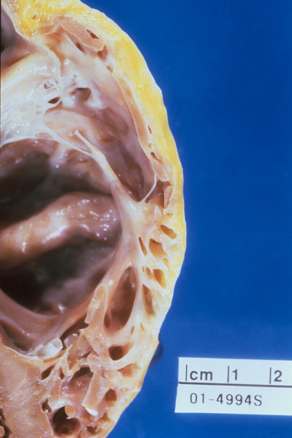
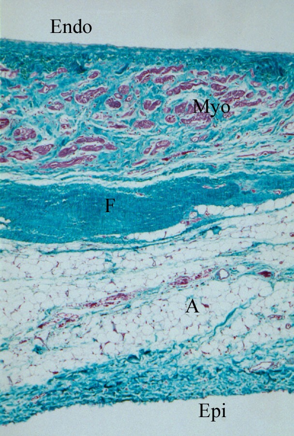
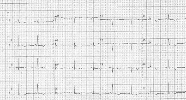
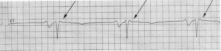
Arrhythmogenic right ventricular cardiomyopathy/dysplasia (ARVC/D) is characterized by the patchy replacement of myocardium by fatty or fibrofatty tissue. These changes lead to structural abnormalities including right ventricular enlargement and wall motion abnormalities that can be detected by echocardiography, angiography, and cine MRI. ARVC/D is a genetically heterogeneous disorder, since it has been linked to several chromosomal loci. Myocarditis may also be a contributing etiological factor. Patients are typically diagnosed during adolescence or young adulthood. Presenting symptoms are generally related to ventricular arrhythmias. Concern for the risk of sudden cardiac death may lead to the implantation of an intracardiac defibrillator. An ongoing multicenter international registry should further our understanding of this disease.
Figures
References
-
- McKoy G, Protonotarios N, Crosby JM, et al. Identification of a deletion in plakoglobin in arrhythmogenic right ventricular cardiomyopathy with palmoplantar keratoderma and woolly hair (Naxos disease) Lancet. 2000;355:2119–2124. - PubMed
-
- Tiso C, Stephan DA, Nava A, et al. Identification of mutations in the cardiac ryanodine receptor gene in families affected with arrhythmogenic right ventircular cardiomyopathy type 2 (ARVD2) Hum Mol Genet. 2001;10:189–194. - PubMed
-
- Corrado D, Thiene G, Nava A, et al. Sudden death in young competitive athletes: clinicopathologic correlations in 22 cases. Am J Medicine. 1990;89:588–596. - PubMed
-
- Marcus FI. Arrhythmogenic right ventricular dysplasia. Cardiac Electrophys Rev. 1999;3:205–206.
LinkOut - more resources
Full Text Sources