Review
. 2006 Nov;326(2):347-59.
doi: 10.1007/s00441-006-0276-3. Epub 2006 Aug 31.

Hair cell ribbon synapses

Affiliations
Review

Hair cell ribbon synapses

Tobias Moser et al. Cell Tissue Res. 2006 Nov.

Abstract

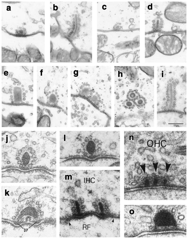
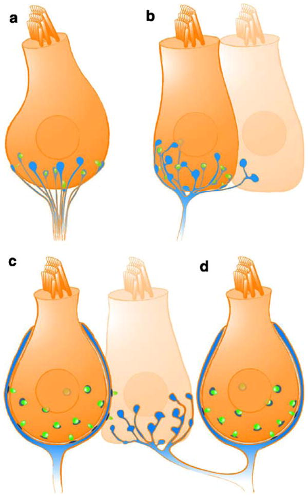
Hearing and balance rely on the faithful synaptic coding of mechanical input by the auditory and vestibular hair cells of the inner ear. Mechanical deflection of their stereocilia causes the opening of mechanosensitive channels, resulting in hair cell depolarization, which controls the release of glutamate at ribbon-type synapses. Hair cells have a compact shape with strong polarity. Mechanoelectrical transduction and active membrane turnover associated with stereociliar renewal dominate the apical compartment. Transmitter release occurs at several active zones along the basolateral membrane. The astonishing capability of the hair cell ribbon synapse for temporally precise and reliable sensory coding has been the subject of intense investigation over the past few years. This research has been facilitated by the excellent experimental accessibility of the hair cell. For the same reason, the hair cell serves as an important model for studying presynaptic Ca(2+) signaling and stimulus-secretion coupling. In addition to common principles, hair cell synapses differ in their anatomical and functional properties among species, among the auditory and vestibular organs, and among hair cell positions within the organ. Here, we briefly review synaptic morphology and connectivity and then focus on stimulus-secretion coupling at hair cell synapses.


Figures

Fig. 1
Fig. 1
Electron micrographs of ribbon synapses from hair cells. ai Ribbons from type I and type II vestibular hair cells in the adult chinchilla crista ampullaris (from Lysakowski and Goldberg 1997, J. Comp. Neurol. vol. 491, pp. 438–439, modified with permission from John Wiley and Sons, Inc.). Type I hair cells generally have small spherical ribbons, regardless of their location within the sensory epithelium (central, a, b; peripheral, c, d). The elongated ribbon from a central type I hair cell in b is an exception in the chinchilla, although much more common in the monkey (Engstrom et al. 1972). Type II hair cells, particularly those in the central part of the sensory epithelium, where sensitivity is highest, show a large variation in shapes and sizes. They can be large and hollow (e, g), spheroid (f), and elongated and plate-like (i), extending through 10–12 ultrathin sections. They can occur singly (e, f, i) or in clusters (g, h). Ribbons in type II hair cells in the peripheral part of the epithelium tend to be small and spheroid (similar to the peripheral type I hair cell ribbons in c, d). jo Ribbons from cochlear hair cells in diverse species. Inner hair cells (jm, IHC) have variable ribbon shapes, whereas outer hair cell (OHC) ribbons tend to be elongated rods (n, o), although the ribbon in o is a less common spheroid ribbon. j, k Middle turn of guinea pig cochlea, inner hair cells (DP dense plaques found presynaptically); modified from Saito (1980) J. Ultrastructural Research, vol. 71, p. 225, with permission from Elsevier. l Cat cochlea, tonotopic location approximately 2.4 kHz, modified from Liberman (1980) Hear. Res., vol. 3, p. 56, with permission from Elsevier. m, n Cat cochlea; inner hair cell (IHC) at 0.5 kHz, and outer hair cell (OHC), respectively, probably from an apical turn, given the multiple ribbons (RF radial fiber afferent, arrows indicate extent of synaptic plaque) arrowheads docked vehicles; modified with permission from Liberman et al. (1990) J. Comp. Neurol. vol. 301, p. 447, with permission from John Wiley and Sons, Inc. (o) Taken from macaque cochlea, outer hair cell, location unknown; from Kimura (1984), modified with permission from Friedmann and Ballantyne (1984) Ultrastructural Altas of the Inner Ear, Taylor and Francis Publishers
Fig. 2
Fig. 2
Afferent synaptic connectivity of hair cells. Four different types of afferent connections are found in the auditory and/or vestibular system. For simplification, all other cellular elements (e.g., efferent contacts and supporting cells) have been ignored. a Several unbranched processes, each forming one synapse (blue fiber and postsynaptic density, green synaptic ribbon) with one hair cell, as is typical for mammalian cochlear inner hair cells. b Branched process innervating two hair cells. Each branch collects input from one or more ribbon synapses. c Calyx-type postsynaptic ending enclosing a type I vestibular hair cell and forming several ribbon synapses with the enclosed hair cell in addition to some synapses on its outer surface with a neighboring type II hair cell. Such “outer face” synapses are more common in the central part of the sensory epithelium. d A dimorphic fiber that forms a calyx synapse with a type I hair cell and bouton synapses with type II hair cells
Fig. 3
Fig. 3
Contrasting views of Ca2+ signaling at hair cell active zones. a Modeling of active zone Ca2+ concentration for frog saccular hair cells; from Roberts (1994), reprinted with permission from the Journal of Neuroscience. The model was based on measurements of voltage-gated Ca2+ entry, the accompanying activation of BK current in frog saccular hair cells, and an estimation of cytosolic Ca2+ buffering capacity. Ca2+ microdomains soak the entire active zone with some locally active maxima. b Representation of the inner hair cell active zone seen from the hair cell cytosolic side with the ribbon (medium gray ellipse projection) removed (from Brandt et al. 2005; reprinted with permission from the Journal of Neuroscience). Synaptic vesicles (gray spheres) preferentially dock to the plasma membrane (transparent) opposite the postsynaptic density (light gray). Approximately 80 Ca2+ channels (black dots) were pseudo-randomly scattered at the active zone with a higher density underneath the ribbon (dark gray ellipsoid) than for the rest of the active zone (light gray ellipsoid postsynaptic density). Synaptic vesicles were placed in a more regular array according to electron microscopic findings (c.f. Fig. 1). Domains of elevated [Ca2+] are indicated in black. In the case of weak stimulation, only a few channels open and drive exocytosis of “their” vesicles, whereas an overlap of Ca2+ domains is expected for saturating stimulation

Similar articles

Cited by

References

    1. Art JJ, Fettiplace R. Variation of membrane properties in hair cells isolated from the turtle cochlea. J Physiol. 1987;385:207–242. - PMC - PubMed
    1. Art JJ, Crawford AC, Fettiplace R. Electrical resonance and membrane currents in turtle cochlear hair cells. Hear Res. 1986;22:31–36. - PubMed
    1. Augustine GJ. How does calcium trigger neurotransmitter release? Curr Opin Neurobiol. 2001;11:320–326. - PubMed
    1. Augustine GJ, Adler EM, Charlton MP. The calcium signal for transmitter secretion from presynaptic nerve terminals. Ann N Y Acad Sci. 1991;635:365–381. - PubMed
    1. Augustine GJ, Santamaria F, Tanaka K. Local calcium signaling in neurons. Neuron. 2003;40:331–346. - PubMed

Publication types

MeSH terms

LinkOut - more resources