

Evaluation of ultrasound lung comets by hand-held echocardiography
- PMID: 16945139
- PMCID: PMC1569440
- DOI: 10.1186/1476-7120-4-34
Evaluation of ultrasound lung comets by hand-held echocardiography
Abstract
Background: Ultrasound lung comets (ULCs) are a clinically useful sign of extravascular lung water. They require very limited technology (2 D-echo), and a short learning curve. The aim of the present study is to compare ULCs information obtained by experienced echocardiologists using a full feature echocardiographic platform and by inexperienced sonographers using a hand-held echocardiography system.
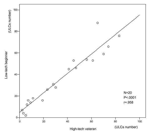
Methods: 20 consecutive in-hospital patients underwent, within 15' and in random order, 2 ultrasound examinations for ULCs by 2 observers with different specific expertise and different technology: 1) "high-tech veteran": ULCs assessment with full feature echocardiographic platform (HP Sonos 7500 Philips Medical Systems, Andover, MA, USA) by a trained echocardiologist, with > or =2 years expertise in ULCs assessment and accredited by the European Association of Echocardiography; 2) and a "low-tech beginner": ULCs assessment with hand-held echocardiography (Optigo; Philips, Andover, MA) by an echocardiographer with very limited (30') dedicated training on ULCs assessment. In each patient, ULC score was obtained by summing the number of comets from each of the scanning spaces in the anterior right and left hemithorax, from the second to the fifth intercostal space.
Results: There was a significant, tight correlation (r = .958, p < 0.001) between the 2 observations in the same patient by "high-tech veteran" and "low-tech beginner".
Conclusion: ULCs are equally reliable in the hands of highly experienced echocardiologists using full feature echocardiographic platforms and in the hands of absolute beginners with miniaturized, compact, and battery-equipped echocardiographic systems. From the technological and expertise viewpoint, ULCs are the "kindergarten" of echocardiography, ideally suited for bedside evaluation of patients with both known or suspected heart failure.
Figures


References
-
- Swedberg K, Cleland J, Dargie H, Drexler H, Follath F, Komajda M, Tavazzi L, Smiseth OA, Gavazzi A, Haverich A, Hoes A, Jaarsma T, Korewicki J, Levy S, Linde C, Lopez-Sendon JL, Nieminen MS, Pierard L, Remme WJ. Task Force for the Diagnosis and Treatment of Chronic Heart Failure of the European Society of Cardiology. Guidelines for the diagnosis and treatment of chronic heart failure: executive summary (update 2005): The Task Force for the Diagnosis and Treatment of Chronic Heart Failure of the European Society of Cardiology. Eur Heart J. 2005;26:1115–1140. doi: 10.1093/eurheartj/ehi166. - DOI - PubMed
-
- Halperin B, Feeley T, Mihm F, et al. Evaluation of the portable chest roentgenogram for quantitating extravascular lung water in critically ill adults. Chest. 1985;88:649–652. - PubMed
-
- Eisenberg PR, Hansbrough JR, Anderson D, et al. A prospective study of lung water measurements during patient management in an intensive care unit. Am Rev Respir Dis. 1987;136:662–668. - PubMed
-
- Targhetta R, Chavagneaux R, Bourgeois JM, et al. Sonographic approach to diagnosing pulmonary consolidation. J Ultrasound Med. 1992;11:667–672. - PubMed
-
- Lichtenstein D, Meziere G, Biderman P, Gepner A, Barre O. The comet-tail artifact. An ultrasound sign of alveolar-interstitial syndrome. Am J Respir Crit Care Med. 1997;156:1640–1646. - PubMed
Publication types
MeSH terms
LinkOut - more resources
Full Text Sources
Research Materials
Miscellaneous
