

Familial ALS-superoxide dismutases associate with mitochondria and shift their redox potentials
- PMID: 16945901
- PMCID: PMC1557633
- DOI: 10.1073/pnas.0605814103
Familial ALS-superoxide dismutases associate with mitochondria and shift their redox potentials
Abstract
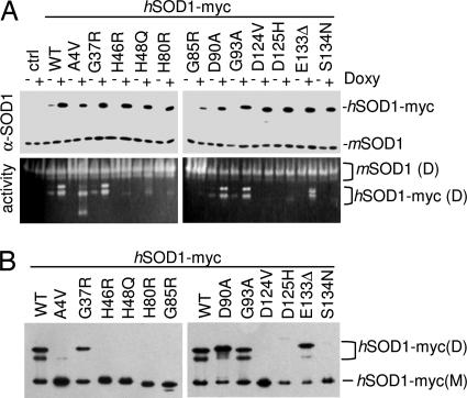
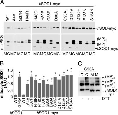
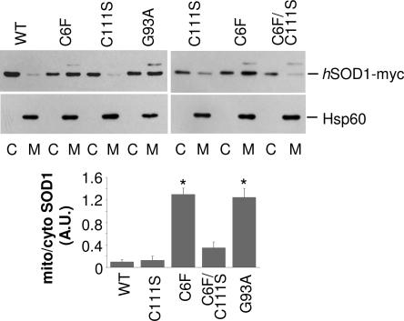
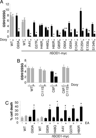
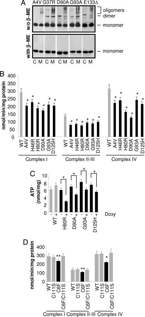
Recent studies suggest that the toxicity of familial amyotrophic lateral sclerosis mutant Cu, Zn superoxide dismutase (SOD1) arises from its selective recruitment to mitochondria. Here we demonstrate that each of 12 different familial ALS-mutant SOD1s with widely differing biophysical properties are associated with mitochondria of motoneuronal cells to a much greater extent than wild-type SOD1, and that this effect may depend on the oxidation of Cys residues. We demonstrate further that mutant SOD1 proteins associated with the mitochondria tend to form cross-linked oligomers and that their presence causes a shift in the redox state of these organelles and results in impairment of respiratory complexes. The observation that such a diverse set of mutant SOD1 proteins behave so similarly in mitochondria of motoneuronal cells and so differently from wild-type SOD1 suggests that this behavior may explain the toxicity of ALS-mutant SOD1 proteins, which causes motor neurons to die.
Conflict of interest statement
Conflict of interest statement: No conflicts declared.
Figures
Similar articles
-
Human Cu/Zn superoxide dismutase (SOD1) overexpression in mice causes mitochondrial vacuolization, axonal degeneration, and premature motoneuron death and accelerates motoneuron disease in mice expressing a familial amyotrophic lateral sclerosis mutant SOD1.Neurobiol Dis. 2000 Dec;7(6 Pt B):623-43. doi: 10.1006/nbdi.2000.0299. Neurobiol Dis. 2000. PMID: 11114261
-
Mutant SOD1 in neuronal mitochondria causes toxicity and mitochondrial dynamics abnormalities.Hum Mol Genet. 2009 Dec 1;18(23):4552-64. doi: 10.1093/hmg/ddp421. Epub 2009 Sep 24. Hum Mol Genet. 2009. PMID: 19779023 Free PMC article.
-
Cell culture models to investigate the selective vulnerability of motoneuronal mitochondria to familial ALS-linked G93ASOD1.Eur J Neurosci. 2006 Jul;24(2):387-99. doi: 10.1111/j.1460-9568.2006.04922.x. Eur J Neurosci. 2006. PMID: 16903849
-
Mitochondrial dysfunction in familial amyotrophic lateral sclerosis.J Bioenerg Biomembr. 2011 Dec;43(6):587-92. doi: 10.1007/s10863-011-9393-0. J Bioenerg Biomembr. 2011. PMID: 22072073 Review.
-
Transgenic mouse model for familial amyotrophic lateral sclerosis with superoxide dismutase-1 mutation.Neuropathology. 2001 Mar;21(1):82-92. doi: 10.1046/j.1440-1789.2001.00361.x. Neuropathology. 2001. PMID: 11304046 Review.
Cited by
-
Antioxidant Alternatives in the Treatment of Amyotrophic Lateral Sclerosis: A Comprehensive Review.Front Physiol. 2020 Feb 6;11:63. doi: 10.3389/fphys.2020.00063. eCollection 2020. Front Physiol. 2020. PMID: 32116773 Free PMC article. Review.
-
A fruitful endeavor: modeling ALS in the fruit fly.Brain Res. 2015 May 14;1607:47-74. doi: 10.1016/j.brainres.2014.09.064. Epub 2014 Oct 5. Brain Res. 2015. PMID: 25289585 Free PMC article. Review.
-
Impaired Autophagy and Defective Mitochondrial Function: Converging Paths on the Road to Motor Neuron Degeneration.Front Cell Neurosci. 2016 Mar 3;10:44. doi: 10.3389/fncel.2016.00044. eCollection 2016. Front Cell Neurosci. 2016. PMID: 26973461 Free PMC article. Review.
-
New Insights into Oxidative Stress and Inflammatory Response in Neurodegenerative Diseases.Int J Mol Sci. 2024 Feb 26;25(5):2698. doi: 10.3390/ijms25052698. Int J Mol Sci. 2024. PMID: 38473944 Free PMC article. Review.
-
Differential effects of phytotherapic preparations in the hSOD1 Drosophila melanogaster model of ALS.Sci Rep. 2017 Jan 19;7:41059. doi: 10.1038/srep41059. Sci Rep. 2017. PMID: 28102336 Free PMC article.
References
-
- Valentine JS, Doucette PA, Potter SZ. Annu Rev Biochem. 2005;74:563–593. - PubMed
-
- Hayward LJ, Rodriguez JA, Kim JW, Tiwari A, Goto JJ, Cabelli DE, Valentine JS, Brown RH., Jr J Biol Chem. 2002;277:15923–15931. - PubMed
-
- Rodriguez JA, Valentine JS, Eggers DK, Roe JA, Tiwari A, Brown RH, Jr, Hayward LJ. J Biol Chem. 2002;277:15932–15937. - PubMed
Publication types
MeSH terms
Substances
Grants and funding
LinkOut - more resources
Full Text Sources
Medical
Miscellaneous
