. 2006 Sep 12;103(37):13676-81.
doi: 10.1073/pnas.0606247103. Epub 2006 Aug 31.

NikR-operator complex structure and the mechanism of repressor activation by metal ions

Affiliations

NikR-operator complex structure and the mechanism of repressor activation by metal ions

Eric R Schreiter et al. Proc Natl Acad Sci U S A. .

Abstract

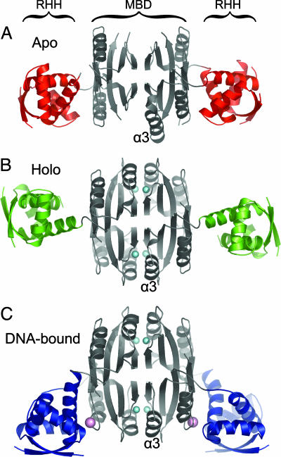
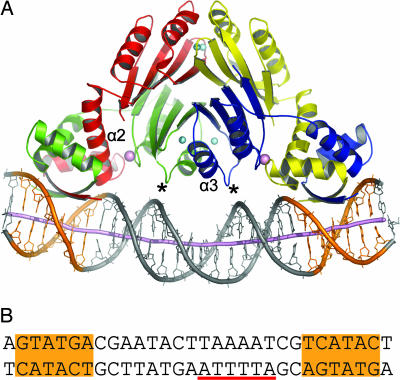
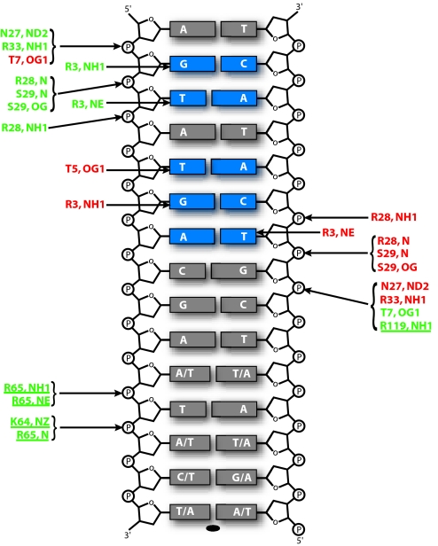
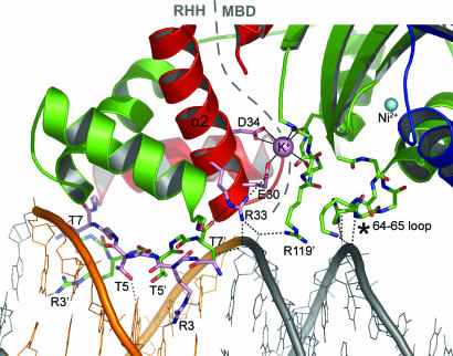
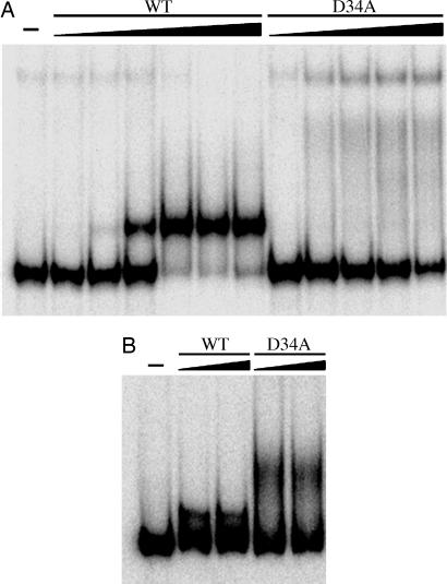
Metal ion homeostasis is critical to the survival of all cells. Regulation of nickel concentrations in Escherichia coli is mediated by the NikR repressor via nickel-induced transcriptional repression of the nickel ABC-type transporter, NikABCDE. Here, we report two crystal structures of nickel-activated E. coli NikR, the isolated repressor at 2.1 A resolution and in a complex with its operator DNA sequence from the nik promoter at 3.1 A resolution. Along with the previously published structure of apo-NikR, these structures allow us to evaluate functional proposals for how metal ions activate NikR, delineate the drastic conformational changes required for operator recognition, and describe the formation of a second metal-binding site in the presence of DNA. They also provide a rare set of structural views of a ligand-responsive transcription factor in the unbound, ligand-induced, and DNA-bound states, establishing a model system for the study of ligand-mediated effects on transcription factor function.


Conflict of interest statement

Conflict of interest statement: No conflicts declared.

Figures

Fig. 1.
Fig. 1.
Conformational flexibility of NikR. (A) Apo-NikR tetramer displayed as a ribbon with the MBD colored gray and the RHH domains colored red. (B) Nickel-activated NikR tetramer displayed as in A, except the RHH domains are colored green and nickel ions are shown as cyan spheres. (C) Operator-bound NikR tetramer displayed as in A and B, except the RHH domains are colored blue and potassium ions are shown as pink spheres. Helix α3, which is stabilized upon high-affinity nickel binding, is labeled in each panel. This figure and other structural figures were made by using PyMOL (DeLano Scientific, San Carlos, CA).
Fig. 2.
Fig. 2.
The NikR–operator DNA complex. (A) NikR–DNA complex with the NikR tetramer colored by subunit, and DNA displayed as sticks with a cartoon tube tracing the phosphorus positions. The dyad-symmetric operator half-sites are colored orange, and the DNA helical axis as calculated in CURVES (15) is shown as a purple tube. Nickel and potassium ions are shown as cyan and pink spheres, respectively. Asterisks indicate the MBD loop that contacts DNA. Helix α2, which contributes the conserved secondary metal site ligands E30 and D34, is labeled on the red NikR molecule. Helix α3, which is structurally stabilized upon high-affinity nickel binding, is labeled on the blue NikR molecule. (B) The dsDNA used for NikR cocrystallization, which includes the wild-type NikR operator sequence. Dyad-symmetric operator half-sites are highlighted in orange, and the -10 region of the nik promoter is underscored with red.
Fig. 3.
Fig. 3.
NikR–operator DNA interactions. Schematic representation of polar interactions between NikR and operator DNA. Only half of the operator DNA is shown because the interactions made with the other half are symmetric and equivalent. Base pairs colored blue were shown to abrogate NikR binding when mutated (4). The protein atoms involved in these interactions are colored by protein subunit and correspond to the color scheme in Fig. 2A. Interactions contributed by the MBD are underlined.
Fig. 4.
Fig. 4.
The second metal-binding site. Protein, DNA, and metal ions are displayed as in Fig. 2. A dashed gray line delineates the RHH domain and MBD. Bonds to the potassium ion at the domain interface are shown as solid black lines, and hydrogen bonds as dashed black lines. Portions of NikR contacting DNA or the secondary metal site are shown as sticks, and important sidechains are labeled. An asterisk indicates the MBD loop, containing Lys-64 and Arg-65, which contacts DNA.
Fig. 5.
Fig. 5.
DNA binding in the presence of excess metal. (A) WT NikR (1, 10, 100 pM, 1, 10, and 100 nM) or the D34A mutant (10 pM, 1, 10, 100 nM, and 1 μM) were preincubated with stoichiometric nickel and incubated subsequently with 100-bp nik DNA in the presence of 35 μM NiSO4. The reactions were analyzed on a 7% native gel with 35 μM NiSO4 in the gel and running buffer. (B) WT NikR (0.5 and 1 μM) and D34A NikR (0.5 and 1 μM) were preincubated with stoichiometric nickel and incubated subsequently with 100-bp nik DNA in the presence of 35 μM KCl. The reactions were analyzed on a 7% native gel with 35 μM KCl in the gel and running buffer. In all mobility-shift assay experiments, KCl (100 mM) was present in the binding buffer.

References

    1. Mulrooney SB, Hausinger RP. FEMS Microbiol Rev. 2003;27:239–261. - PubMed
    1. Navarro C, Wu LF, Mandrand-Berthelot MA. Mol Microbiol. 1993;9:1181–1191. - PubMed
    1. De Pina K, Desjardin V, Mandrand-Berthelot MA, Giordano G, Wu LF. J Bacteriol. 1999;181:670–674. - PMC - PubMed
    1. Chivers PT, Sauer RT. J Biol Chem. 2000;275:19735–19741. - PubMed
    1. Contreras M, Thiberge JM, Mandrand-Berthelot MA, Labigne A. Mol Microbiol. 2003;49:947–963. - PubMed

Publication types