. 2006 Sep 12;103(37):13652-7.
doi: 10.1073/pnas.0606140103. Epub 2006 Aug 31.

Purification and molecular cloning of a DNA ADP-ribosylating protein, CARP-1, from the edible clam Meretrix lamarckii

Affiliations

Purification and molecular cloning of a DNA ADP-ribosylating protein, CARP-1, from the edible clam Meretrix lamarckii

Tsuyoshi Nakano et al. Proc Natl Acad Sci U S A. .

Abstract

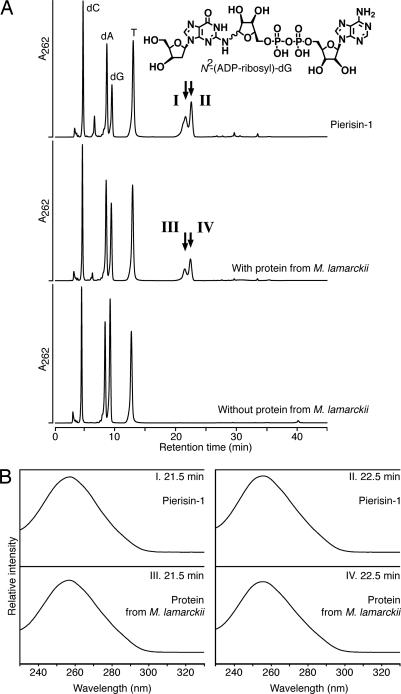
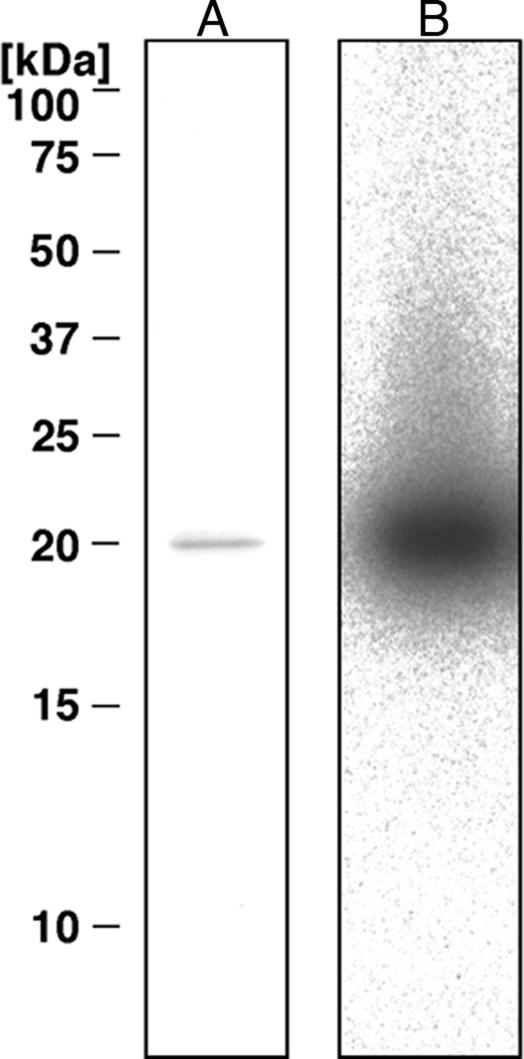
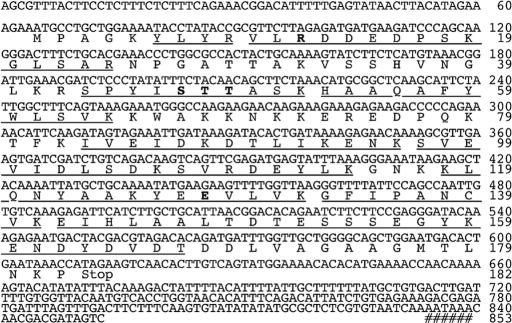
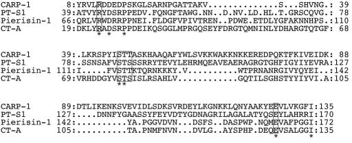
The cabbage butterflies Pieris rapae and Pieris brassicae have unique enzymes, named pierisin-1 and -2, respectively, that catalyze the ADP-ribosylation of guanine residues of DNA, which has been linked with induction of apoptosis and mutation in mammalian cell lines. In the present study, we identified ADP-ribosylation activity targeting DNA in six kinds of edible clam. Similar to our observations with pierisin-1 and -2, crude extracts from the clams Meretrix lamarckii, Ruditapes philippinarum, and Corbicula japonica incubated with calf thymus DNA and beta-NAD resulted in production of N(2)-(ADP-ribos-1-yl)-2'-deoxyguanosine. The DNA ADP-ribosylating protein in the hard clam M. lamarckii, designated as CARP-1, was purified by column chromatography, and its cDNA was cloned. The cDNA encodes a 182-aa protein with a calculated molecular mass of 20,332. The protein synthesized in vitro from the cDNA in a reticulocyte lysate exhibited the same ADP-ribosylating activity as that of purified CARP-1. Neither the nucleotide nor the deduced amino acid sequence of CARP-1 showed homology with pierisin-1 or -2. However, a glutamic acid residue (E128) at the putative NAD-binding site, conserved in all ADP-ribosyltransferases, was found in CARP-1, and replacement of aspartic acid for this glutamic acid resulted in loss of almost all ADP-ribosylating activity. CARP-1 in the culture medium showed no cytotoxicity against HeLa and TMK-1 cells; however, introduction of this protein by electroporation induced apoptosis in these cells. The finding of clam ADP-ribosylating protein targeting guanine residues in DNA could offer new insights into the biological significance of ADP-ribosylation of DNA.


Conflict of interest statement

Conflict of interest statement: No conflicts declared.

Figures

Fig. 1.
Fig. 1.
Detection of ADP-ribosylated DNA adducts formed with crude extracts of clams. Calf thymus DNA was reacted with 32P-NAD in the presence of 1 μg of crude extract protein. The DNA samples were spotted on TLC sheets with and without nuclease digestion, which were developed and subjected to autoradiography. Sample 1, no protein; sample 2, pierisin-1 (10 pg, positive control); sample 3, M. lamarckii; sample 4, R. philippinarum; sample 5, C. japonica; sample 6, My. galloprovincialis. The arrow indicates the Rf value of 0.05. +, with nuclease digestion; −, without nuclease digestion.
Fig. 2.
Fig. 2.
Analysis of reaction products formed by DNA, clam protein, and β-NAD. (A) HPLC elution patterns of hydrolysate of DNA incubated with pierisin-1 (Top), with partially purified protein from M. lamarckii (Middle), or without protein (Bottom). These DNA samples were enzymatically hydrolyzed to deoxyribonucleosides and injected into a Develosil RPAQUEOUS column, and the eluate was monitored by measuring its UV absorbance at 262 nm. Arrows indicate the peaks coincident with N2-(ADP-ribos-1-yl)-2′-deoxyguanosine. (B) UV absorption spectra with a photodiode array detector of the compounds (IIV) in the peak fractions at retention times of 21.5 and 22.5 min presented in A.
Fig. 3.
Fig. 3.
SDS/PAGE of purified CARP-1. One microgram of purified protein was separated by SDS/PAGE by using 15% gels, which were stained with Coomassie brilliant blue R-250 (A) or assessed for activity by using the activity gel assay (B).
Fig. 4.
Fig. 4.
Nucleotide and deduced amino acid sequences of the CARP-1 cDNA determined by PCR direct sequencing. The ORF is located between nucleotides 65 and 613. All determined internal amino acid sequences of the purified CARP-1 protein are indicated by underlining. A possible polyadenylation signal is indicated by #. Possible conserved amino acids are shown in bold type.
Fig. 5.
Fig. 5.
Alignment of the deduced amino acid sequence of the homologous region of CARP-1 with pierisin-1 and ADP-ribosylating toxins. PT-S1, the S1 subunit of PT (GenBank accession no. P04977); pierisin-1 (9); CT-A, the A subunit of CT (GenBank accession no. 1001196A). The conserved arginine, Ser–Thr–Ser/Thr motif, and glutamic acid residues are boxed. Completely conserved amino acids are marked by asterisks.

References

    1. Sugimura T. Prog Nucleic Acid Res Mol Biol. 1973;13:127–151. - PubMed
    1. Seman M, Adriouch S, Haag F, Koch-Nolte F. Curr Med Chem. 2004;11:857–872. - PubMed
    1. Krueger KM, Barbieri JT. Clin Microbiol Rev. 1995;8:34–47. - PMC - PubMed
    1. Okazaki IJ, Moss J. Annu Rev Nutr. 1999;19:485–509. - PubMed
    1. Zhao Z, Gruszczynska-Biegala J, Zolkiewska A. Biochem J. 2005;385:309–317. - PMC - PubMed

Publication types

Associated data