. 2006 Sep 12;103(37):13837-42.
doi: 10.1073/pnas.0600843103. Epub 2006 Aug 31.

Leishmania disease development depends on the presence of apoptotic promastigotes in the virulent inoculum

Affiliations

Leishmania disease development depends on the presence of apoptotic promastigotes in the virulent inoculum

Ger van Zandbergen et al. Proc Natl Acad Sci U S A. .

Erratum in

  • Proc Natl Acad Sci U S A. 2006 Oct 31;103(44):16615

Abstract

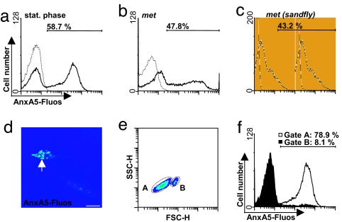
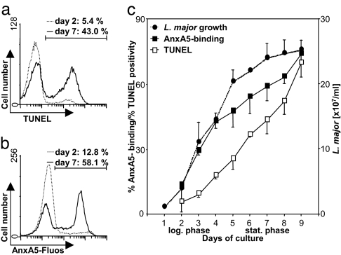
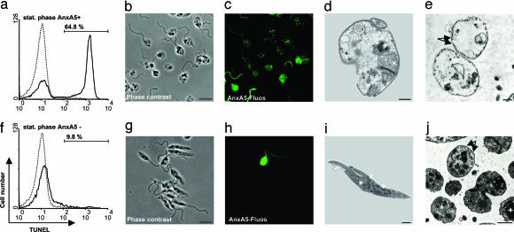
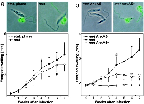
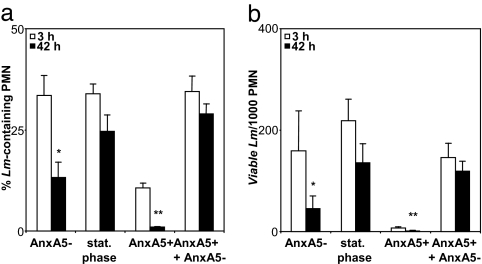
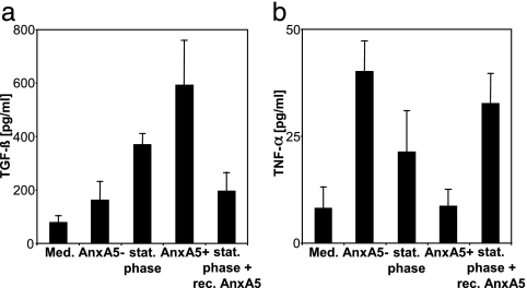
The obligate intracellular pathogen Leishmania major survives and multiplies in professional phagocytes. The evasion strategy to circumvent killing by host phagocytes and establish a productive infection is poorly understood. Here we report that the virulent inoculum of Leishmania promastigotes contains a high ratio of annexin A5-binding apoptotic parasites. This subpopulation of parasites is characterized by a round body shape, a swollen kinetoplast, nuclear condensation, and a lack of multiplication and represents dying or already dead parasites. After depleting the apoptotic parasites from a virulent population, Leishmania do not survive in phagocytes in vitro and lose their disease-inducing ability in vivo. TGF-beta induced by apoptotic parasites is likely to mediate the silencing of phagocytes and lead to survival of infectious Leishmania populations. The data demonstrate that apoptotic promastigotes, in an altruistic way, enable the intracellular survival of the viable parasites.


Conflict of interest statement

Conflict of interest statement: No conflicts declared.

Figures

Fig. 1.
Fig. 1.
AnxA5 binding to L. major promastigotes. Populations of L. major promastigotes were stained with AnxA5-Fluos. Flow cytometry histogram profiles of stat. phase promastigotes (black line, a) and of stat. phase-derived metacyclic promastigotes (met, black line, b). The dotted lines (a and b) show the control staining in the absence of Ca2+. (c) Metacyclic promastigotes derived from P. duboscqi sandflies (black line) as described (22). The dotted line shows the unstained control. (d) Confocal micrograph (0.15-μM slice) of a promastigote-stained positive with AnxA5 (arrow) and an AnxA5− promastigote. (Scale bar, 5 μm.) (e) Flow cytometry densitoblot showing forward scatter (FSC-H) and sideward scatter (SSC-H) analysis of the stat. phase promastigotes. (f) AnxA5-Fluos binding of the population A in e (white histogram) and population B in e (filled histogram).
Fig. 2.
Fig. 2.
TUNEL and AnxA5 staining of L. major promastigotes in the log. and stat. growth phases. Parasites were stained with the TUNEL method (a) or by using AnxA5-Fluos (b) and analyzed with flow cytometry. The histogram profiles of early log. phase (dotted line) and stat. phase (black line) parasites are shown. (c) Parasite growth (dotted line) and percentage of AnxA5+ (black line, filled squares) and TUNEL+ promastigotes (black line, open squares) during the course of in vitro parasite growth. The histograms are representative for four independent experiments. Data of four independent experiments are depicted as mean ± SD.
Fig. 3.
Fig. 3.
Separation of AnxA5+ promastigotes from AnxA5− parasites. In vitro cultured stat. phase promastigotes were separated into AnxA5+ (stat. phase AnxA5+) and AnxA5− (stat. phase AnxA5−) populations by using AnxA5-based MACS. Parasites were stained with the TUNEL method (a and f) and analyzed with flow cytometry. The histogram profiles of AnxA5+ (black line, a) and AnxA5− (black line, f) parasites are shown. The dotted lines show the TUNEL control staining, with fluorescent label added without addition of terminal deoxynucleotidyl transferase. Phase-contrast micrograph of AnxA5+ (b) and AnxA5− (g) populations. (Scale bar, 5 μm.) AnxA5-Fluos-staining of AnxA5+ (c) and AnxA5− (h) populations. Electron micrographs of longitudinal parasite sections (d and i) or transverse parasite sections (e and j) of purified AnxA5+ (d and e) and purified AnxA5− (i and j) parasites showing the mitochondrion/kinetoplast structure (K) at the end of the flagella pocket, nuclear structure (+), cytoplasmic granular-like structures (∗), and tubular organization (arrows). Micrographs and electron micrographs are representative of four independent experiments.
Fig. 4.
Fig. 4.
PS-dependent disease development in mice. (a) AnxA5-Fluos staining combined with phase-contrast micrographs are depicted of stat. phase promastigotes (stat. phase, Upper Left) and stat. phase-derived metacyclic L. major promastigotes (met, Upper Right). (Scale bar, 5 μm.) Mice were infected with 1 × 10⁶ stat. phase promastigotes (stat. phase, open circles) or stat. phase-derived metacyclic L. major promastigotes (met, filled circles). Disease development was assessed by measuring the swelling of the infected footpad. (b) Micrographs (as in a) are depicted of metacyclic-derived AnxA5− (met AnxA5 −, Upper Left) and metacyclic derived AnxA5+ (met AnxA5 +, Upper Right). (Scale bar, 5 μm.) Mice were infected with 1 × 10⁶ metacyclic L. major promastigotes (met, filled circles) as well as from this population-derived AnxA5− positive (met AnxA5 +, open squares) or AnxA5− (met AnxA5−, open circles) parasites. Data (mean ± SD) are from one representative experiment with eight mice per group of three experiments performed. # indicates onset of footpad necrosis, and ∗∗ indicates P < 0.0005.
Fig. 5.
Fig. 5.
Intracellular presence and survival L. major promastigote populations in human PMN. PMN were coincubated with stat. phase, stat. phase-derived purified AnxA5− or AnxA5+, or a 1:1 mixture of AnxA5+ and AnxA5− promastigotes. (a) The percentage of PMN-containing intracellular parasites was determined 3 and 42 h after coincubation using microscopical analysis of Giemsa-stained preparates. (b) Intraneutrophilic parasite survival 3 and 42 h after coincubation was assessed by using end-point titration and is depicted as the number of parasites per 1,000 PMN. Data show mean ± SD of three independent experiments. ∗, P < 0.05; ∗∗, P < 0.005.
Fig. 6.
Fig. 6.
Cytokine production by human phagocytes after exposure to apoptotic and nonapoptotic L. major promastigotes. PMN (a and b) were cultured for 18 h in medium alone with stat. phase promastigotes, stat. phase-derived purified AnxA5− or AnxA5+ promastigotes, or with stat. phase promastigotes preincubated with 5 μg of recombinant annexin-V (Responsif; stat. phase + recAnxA5) at a parasite/PMN ratio of 5:1. The TGF-β (a) and TNF-α (b) content of the supernatants was measured by using an ELISA. Data show mean ± SD of four independent experiments.

References

    1. da Silva R, Sacks DL. Infect Immun. 1987;55:2802–2806. - PMC - PubMed
    1. Sacks DL, Hieny S, Sher A. J Immunol. 1985;135:564–569. - PubMed
    1. Spath GF, Epstein L, Leader B, Singer SM, Avila HA, Turco SJ, Beverley SM. Proc Natl Acad Sci USA. 2000;97:9258–9263. - PMC - PubMed
    1. Sunderkotter C, Kunz M, Steinbrink K, Meinardus-Hager G, Goebeler M, Bildau H, Sorg C. J Immunol. 1993;151:4891–4901. - PubMed
    1. Laufs H, Müller K, Fleischer J, Reiling N, Jahnke N, Jensenius JC, Solbach W, Laskay T. Infect Immun. 2002;70:826–835. - PMC - PubMed

Publication types

Substances