. 2006;34(16):4537-45.
doi: 10.1093/nar/gkl634. Epub 2006 Aug 31.

Tobacco plastid ribosomal protein S18 is essential for cell survival

Affiliations

Tobacco plastid ribosomal protein S18 is essential for cell survival

Marcelo Rogalski et al. Nucleic Acids Res. 2006.

Abstract

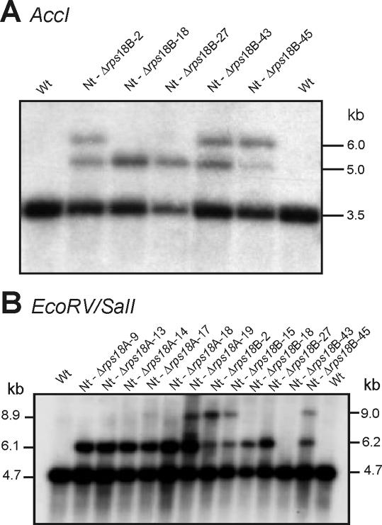
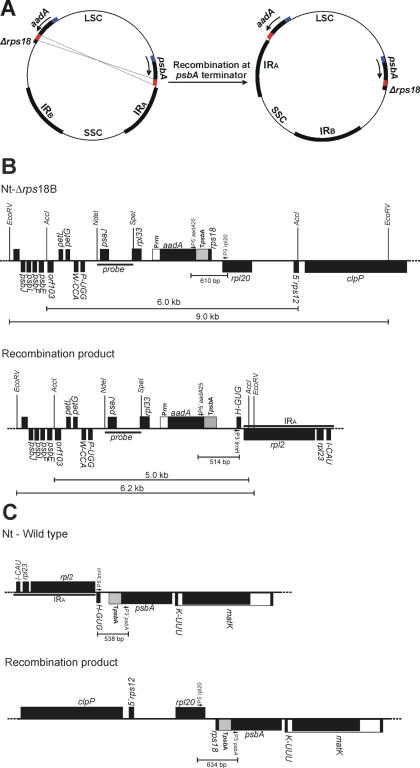
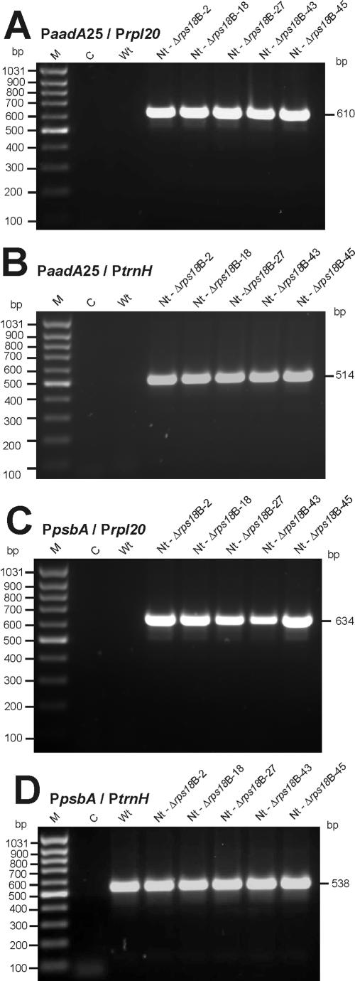
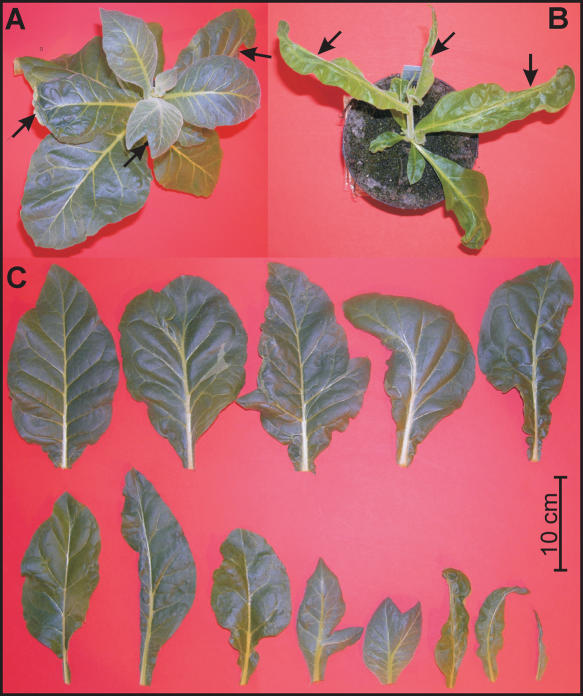
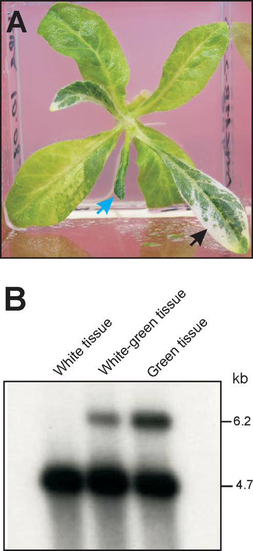
Plastid genomes contain a conserved set of genes most of which are involved in either photosynthesis or gene expression. Among the ribosomal protein genes present in higher plant plastid genomes, rps18 is special in that it is absent from the plastid genomes of several non-green unicellular organisms, including Euglena longa and Toxoplasma gondii. Here we have tested whether the ribosomal protein S18 is required for translation by deleting the rps18 gene from the tobacco plastid genome. We report that, while deletion of the rps18 gene was readily obtained, no homoplasmic Deltarps18 plants or leaf sectors could be isolated. Instead, segregation into homoplasmy led to severe defects in leaf development suggesting that the knockout of rps18 is lethal and the S18 protein is required for cell survival. Our data demonstrate that S18 is indispensable for plastid ribosome function in tobacco and support an essential role for plastid translation in plant development. Moreover, we demonstrate the occurrence of flip-flop recombination on short inverted repeat sequences which generates different isoforms of the transformed plastid genome that differ in the orientation a 70 kb segment in the large single-copy region. However, infrequent occurrence of flip-flop recombination and random segregation of plastid genomes result in the predominant presence of only one of the isoforms in many tissue samples. Implications for the interpretation of chloroplast transformation experiments and vector design are discussed.


Figures

Figure 1
Figure 1
Construction of plastid transformation vectors for disruption of rps18. (A) Physical maps of the rps18 region in the tobacco plastid genome [ptDNA; Ref. (15)]. Genes above the lines are transcribed from the left to the right, genes below the lines are transcribed in the opposite direction. (B) Map of transformation vector pΔrps18A. Note that the spectinomycin resistance gene aadA is driven by the endogenous rps18 promoter. (C) Map of transformation vector pΔrps18B. In this vector, the rRNA operon-derived chimeric Prrn promoter (16) drives the selectable marker gene aadA. Restriction sites used for cloning, RFLP analysis and/or generation of hybridization probes are indicated. Sites lost owing to ligation to heterologous ends are shown in parentheses. The hybridization probe (NdeI/SpeI fragment) is indicated and the expected sizes of hybridizing bands in the two RFLP analyses (Figure 2) is shown below each map. The chloroplast targeting fragment in transformation vectors pΔrps18A and pΔrps18B is marked by dashed lines (NdeI/EcoRI fragment cloned into pUC18).
Figure 2
Figure 2
RFLP analysis of chloroplast transformants obtained with rps18 knockout constructs. Equal amounts of extracted total cellular DNA (5 µg per sample) were digested with the restriction enzymes indicated, separated in 0.8% agarose gels, blotted and hybridized to a radiolabeled restriction fragment derived from cloned tobacco plastid DNA (NdeI/SpeI fragment; Figure 1). (A) RFLP with AccI. (B) RFLP with EcoRV and SalI. The probe detects, in addition to the expected fragments (Figure 1), an unexpected band in most samples which is the product of flip-flop recombination (see text for details and Figures 3 and 4). Fragment sizes of the molecular weight marker are given in kb. Lane Nt-Δrps18B-43 is positive in the AccI RFLP and negative in the EcoRV/SalI RFLP most probably because the leaf sample harvested for DNA extraction for the AccI blot was heteroplasmic, whereas the sample harvested for isolating the DNA for the EcoRV/SalI blot had segregated into virtual homoplasmy for the wild-type genome. Wt: wild type.
Figure 3
Figure 3
Model for flip-flop recombination on the two copies of the psbA 3′-UTR present in the transplastomes. (A) Inversion of a large, 70 kb segment of the large single copy region (LSC) by recombination between the two homologous sequences. IRA and IRB, inverted repeat regions; SSC, small single copy region. (B) Maps of the two different genome conformations in the psaJ/rpl33 region of the plastid genome. Restriction fragments hybridizing to the probe are indicated below each map (cp. Figure 2). (C) Maps of the two different genome conformations in the psbA/trnK/matK region of the plastid genome. PCR primers and expected sizes of amplification products in PCR assays diagnostic for the different genome conformations are also indicated.
Figure 4
Figure 4
Confirmation of flip-flop recombination by PCR. For location and orientation of PCR primers, see Figure 3. M, molecular weight marker; C, buffer control; Wt, wild type control. Sizes of obtained amplification products are indicated at the right in base pair. (A) Test for the expected genome conformation in transplastomic lines (see Figure 3B upper panel). (B) Test for the product of flip-flop recombination in the psaJ/rpl33 region of transplastome (see Figure 3B lower panel). (C) Test for the product of flip-flop recombination in the psbA/trnK/matK region of transplastome (see Figure 3C lower panel). (D) Control amplification of the psbA 3′-UTR from the wild-type genome (see Figure 3C upper panel).
Figure 5
Figure 5
Seed assays confirming heteroplasmy and random genome segregation in rps18 knockout lines. (A) Seed germination in the absence of spectinomycin. (B) Wild-type control on medium with spectinomycin. (C and D) Seeds from two independently generated transplastomic lines germinated in the presence of spectinomycin (C: Nt-Δrps18B-27; D: Nt-Δrps18B-15). In white seedlings, the transplastome has been lost owing to random sorting out of plastid genomes. In contrast, green and variegated seedlings contain the transplastome and hence, express spectinomycin resistance. Approximately 1500 seeds were sown per plate.
Figure 6
Figure 6
Mutant phenotypes of Nt-Δrps18 tobacco plants. (A and B) Leaf abnormalities in greenhouse-grown plants (T0 generation). Arrows point to misshapen leaves that lack parts of the leaf blade. The phenotype is variable and depends on the frequency of somatic segregation towards homoplasmy for the rps18 knockout. (A) Example of a plant with a relatively mild phenotype. (B) Example of a plant with a severe phenotype. (C) Spectrum of leaf phenotypes observed in Nt-Δrps18 plants. Phenotypes range from nearly normal leaves (top row left) to needle-like mutant leaves lacking the entire leaf blade (bottom row right).
Figure 7
Figure 7
Genome segregation in the absence of selection for the transplastome (A) Leaf phenotype in the spectinomycin re-exposure experiments. Plants were initially raised on spectinomycin-containing medium (note blue arrow pointing to a misshapen leaf), then transferred to spectinomycin-free medium for 3 weeks followed by re-exposure to spectinomycin-containing medium. Spectinomycin re-exposure visualizes sectors that have lost the transplastome and, therefore. lack the aadA spectinomycin resistance gene (bleaching sectors indicated by the black arrow). (B) RFLP analysis to confirm the absence of transformed Δrps18 genomes in white sectors of Nt-Δrps18 leaves grown in the absence of spectinomycin and then re-exposed to the drug. The restriction enzyme combination used for the RFLP was EcoRV/SalI (cp. Figure 2B). Of total cellular DNA 1 µg was digested and separated in a 0.8% agarose gel. Whereas green sectors show the 6.2 kb transplastome-specific band, white sectors show only a signal for the 4.7 kb wild-type band.

Similar articles

Cited by

References

    1. Mache R. Chloroplast ribosomal proteins and their genes. Plant Sci. 1990;72:1–12.
    1. Sugiura M., Hirose T., Sugita M. Evolution and mechanism of translation in chloroplasts. Annu. Rev. Genet. 1998;32:437–459. - PubMed
    1. Harris E.H., Boynton J.E., Gillham N.W. Chloroplast ribosomes and protein synthesis. Microbiol. Rev. 1994;58:700–754. - PMC - PubMed
    1. Gockel G., Hachtel W., Baier S., Fliss C., Henke M. Genes for components of the chloroplast translational apparatus are conserved in the reduced 73-kb plastid DNA of the nonphotosynthetic euglenoid flagellate Astasia longa. Curr. Genet. 1994;26:256–262. - PubMed
    1. Wilson R.J.M., Denny P.W., Preiser P.R., Rangachari K., Roberts K., Roy A., Whyte A., Strath M., Moore D.J., Moore P.W., et al. Complete gene map of the plastid-like DNA of the malaria parasite Plasmodium falciparum. J. Mol. Biol. 1996;261:155–172. - PubMed

Publication types