. 2006;34(16):4495-505.
doi: 10.1093/nar/gkl614. Epub 2006 Aug 31.

CAG*CTG repeat instability in cultured human astrocytes

Affiliations

CAG*CTG repeat instability in cultured human astrocytes

Brian T Farrell et al. Nucleic Acids Res. 2006.

Abstract

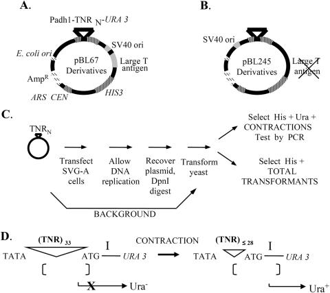
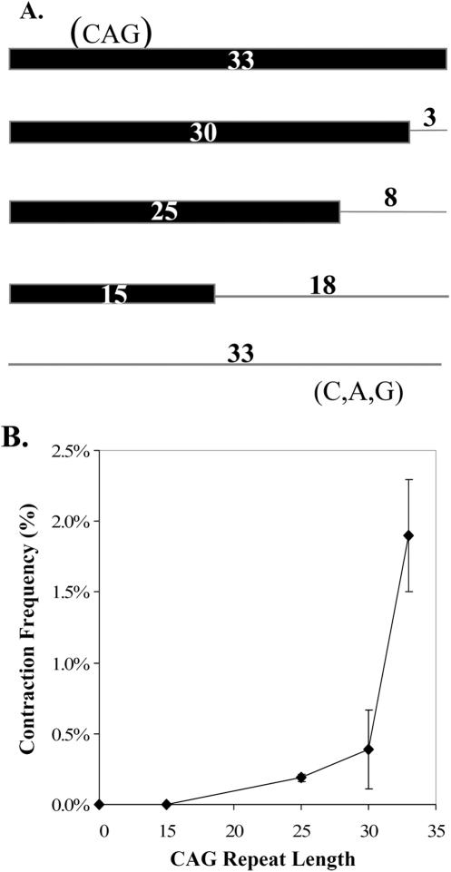
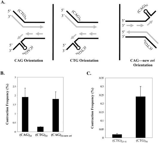
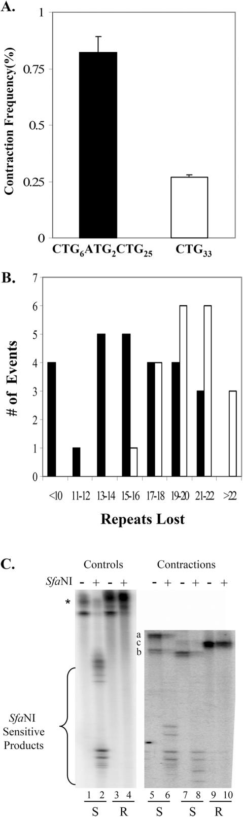
Cells of the central nervous system (CNS) are prone to the devastating consequences of trinucleotide repeat (TNR) expansion. Some CNS cells, including astrocytes, show substantial TNR instability in affected individuals. Since astrocyte enrichment occurs in brain regions sensitive to neurodegeneration and somatic TNR instability, immortalized SVG-A astrocytes were used as an ex vivo model to mimic TNR mutagenesis. Cultured astrocytes produced frequent (up to 2%) CAG.CTG contractions in a sequence-specific fashion, and an apparent threshold for instability was observed between 25 and 33 repeats. These results suggest that cultured astrocytes recapitulate key features of TNR mutagenesis. Furthermore, contractions were influenced by DNA replication through the repeat, suggesting that instability can arise by replication-based mechanisms in these cells. This is a crucial mechanistic point, since astrocytes in the CNS retain proliferative capacity throughout life and could be vulnerable to replication-mediated TNR instability. The presence of interruptions led to smaller but more frequent contractions, compared to a pure repeat, and the interruptions were sometimes deleted to form a perfect tract. In summary, we suggest that CAG.CTG repeat instability in cultured astrocytes is dynamic and replication-driven, suggesting that TNR mutagenesis may be influenced by the proliferative capacity of key CNS cells.


Figures

Figure 1
Figure 1
Shuttle vector plasmids and assay outline. Details are provided in Materials and Methods. (A) The shuttle vector pBL67 contains several important genetic elements enabling analysis of TNR instability in cultured cells. Genetic elements tinted gray, such as the SV40 origin of replication, drive plasmid replication in SVG-A cells. A striped-pattern indicates yeast genetic elements for determination of contraction frequency by selection in Saccharomyces cerevisiae. Dashed bars indicate Escherichia coli elements that enable replication and large-scale preparation of plasmid by plasmid maxi-kit (Qiagen Inc). (B) The vector pBL245 follows the same pattern scheme, the only difference between the two vectors being the absence of the large T antigen and the relocation of the SV40 ori to the opposite side of the repeat tract. (C) Each TNR-containing vector was transfected into SVG-A cells, the cells were cultured for 2–3 days, then the plasmid DNA was isolated and transformed into yeast for analysis. Contraction frequency was calculated by dividing the number of yeast colonies with a contraction by the total number of transformants. Background contraction frequencies were measured as described in Materials and Methods. (D) The selection for contractions in yeast is based on spacing sensitivity of the Schizosaccharomyces pombe adh1 promoter to the distance between the TATA box and the preferred transcription initiation site, labeled ‘I’. Starting TNR lengths of 33 inhibit expression of the URA3 reporter gene and yield a Ura phenotype (36). TNR contractions that remove at least five repeats restore promoter activity and result in a Ura+ phenotype.
Figure 2
Figure 2
Analysis of SVG-A cells and transfected TNR plasmid DNA. (A) SVG-A cells stained with anti-GFAP antibodies (green false color) and counterstained with DAPI (blue false color) at 400× magnification. (B) DpnI resistance assay after recovery of plasmid pBL248 from SVG-A cells and subsequent PCR analysis of contracted alleles after yeast selection. The left panel shows the result of DpnI digestion of stock plasmid used for background determinations (lane 1 = uncut, lane 2 = cut) and plasmid DNA that has been passaged through SVG-A cells (lane 3 = uncut, lanes 4–6 = cut). Sample wells between lanes 2 and 3 and lanes 3 and 4 were intentionally left empty. The right panel shows the analysis of individual contraction events from SVG-A cells. PCR amplification of starting plasmid DNA indicates the size of the 33-repeat parental allele (lane 7). Individual contractions are shown in lanes 8–13. Fragment sizes, stated as repeat units, were deduced by comparison with molecular weight standards (data not shown).
Figure 3
Figure 3
Repeat-length dependence for TNR contractions in SVG-A cells. (A) The top line represents a perfect (CAG)33 allele while the bottom indicates a scrambled (C,A,G)33 allele with no repeating nature (zero repeats). The thick bar indicates the length of the perfect CAG repeat while the thin line shows the number of scrambled C,A, and G triplet equivalents appended to each repeat in order to normalize all constructs to 99 nt. (B) Response of CAG contractions in SVG-A cells to changes in initial tract length. The contraction frequency (Table 1) on the ordinate was derived by subtracting the background contraction frequency from the contraction frequency observed in SVG-A cells. Error bars indicate ±1 SEM.
Figure 4
Figure 4
The repeat orientation relative to the direction of replication influences CAG·CTG contractions in SVG-A cells. (A) Complementary CAG and CTG repeats can differentially affect TNR instability based on the orientation of the repeat relative to the direction of replication. In the left panel, CAG repeats reside on the lagging strand template. In the middle panel, the reverse orientation places the CTG repeats on the lagging template strand. On the right, switching the position of the SV40 ori reverses the direction of replication and swaps the assignment of leading and lagging strand. The direction of replication was deduced from the position of the S40 ori relative to the TNR. (B) Contraction frequency (%) is plotted as given in Table 1 (column 9) for the indicated starting alleles. Error bars indicate ±1 SEM. (C) Contraction frequency (%) is plotted versus CTG repeat length. All values in (B and C) represent the corrected contraction frequency in SVG-A cells.
Figure 5
Figure 5
Influence of interruptions on CTG repeat contractions in SVG-A cells. (A) Corrected contraction frequencies (Table 1, entries 9L and 9M) are plotted for starting tracts of (CTG)6(ATG)2(CTG)25 (filled bar) and for (CTG)33 (unfilled bar). Error bars indicate ±1 SEM. (B) Mutation spectra for (CTG)6(ATG)2(CTG)25 (filled bars) versus (CTG)33 (unfilled bars). The number of observed events is plotted on the ordinate versus the size of the contraction (as determined by PCR) on the abscissa. (C) Representative gel showing results of the SfaNI-resistance assay. PCR products were analyzed on 6% denaturing polyacrylamide gels prior to (odd number lanes) or following (even number lanes) treatment with SfaNI, as described in Materials and Methods. Lanes 1–4 show results with PCR products from control 33-repeat plasmids that are interrupted (lanes 1 and 2) or perfect (lanes 3 and 4). The asterisk indicates the starting position of the 33-repeat uncut and SfaNI-resistant perfect repeat tracts. Lanes 5–10 show results with PCR products from contractions of the (CTG)6(ATG)2(CTG)25 allele in SVG-A cells. The letters a–c indicate the position of the uncut samples in lanes 5, 7 and 9, respectively. The letters below the gel image indicate the assignment of SfaNI sensitivity (S) or resistance (R). To become resistant, both ATG interruptions must be lost.

Similar articles

Cited by

References

    1. Paulson H.L., Fischbeck K.H. Trinucleotide repeats in neurogenetic disorders. Annu. Rev. Neurosci. 1996;19:79–107. - PubMed
    1. Mirkin S.M. Molecular models for repeat expansions. Chemtracts-Biochem. Mol. Biol. 2004;17:639–662.
    1. Pearson C.E., Edamura K.N., Cleary J.D. Repeat instability: mechanisms of dynamic mutations. Nature Rev. Genet. 2005;6:729–742. - PubMed
    1. Kovtun I.V., McMurray C.T. Trinucleotide expansion in haploid germ cells by gap repair. Nature Genet. 2001;27:407–411. - PubMed
    1. Gomes-Pereira M., Fortune M.T., Monckton D.G. Mouse tissue culture models of unstable triplet repeats: in vitro selection for larger alleles, mutational expansion bias and tissue specificity, but no association with cell division rates. Hum. Mol. Genet. 2001;10:845–854. - PubMed

Publication types