

Long-term changes of functional MRI-based brain function, behavioral status, and histopathology after transient focal cerebral ischemia in rats
- PMID: 16946164
- PMCID: PMC2949951
- DOI: 10.1161/01.STR.0000239667.15532.c1
Long-term changes of functional MRI-based brain function, behavioral status, and histopathology after transient focal cerebral ischemia in rats
Abstract
Background and purpose: The relation between recovery of brain function and neurological status after clinical and experimental cerebral ischemia is incompletely characterized. We assessed the evolution of ischemic injury, behavioral status, and brain activity at acute to chronic periods after transient middle cerebral artery occlusion (tMCAO) in rats.
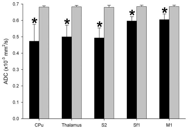
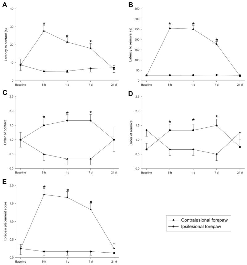
Methods: Male Sprague-Dawley rats were subjected to 20-minute tMCAO (n=10) or sham operation (n=10). Sensorimotor behavioral testing and multimodal (diffusion, perfusion, T2, and functional) MRI, as well as postmortem hematoxylin-eosin staining, were performed before and up to 21 days after tMCAO. MRI and histological parameters were evaluated in 5 regions of interest within the sensorimotor network. Diffusion, perfusion, and T2 lesion volumes were calculated according to previously established viability thresholds.
Results: Diffusion and perfusion lesions were present during occlusion but disappeared completely and permanently within 30 minutes after reperfusion, with no T2 lesions seen. Functional MRI and behavioral deficits did not normalize until 1 and 21 days after tMCAO, respectively. Histology demonstrated selective neuronal cell death at 7 and 21 days after reperfusion.
Conclusions: Twenty-minute tMCAO produced distinct changes on multimodal MRI, histology, and behavioral parameters acutely and chronically. Normal findings on MRI after transient ischemia may not indicate normal tissue status, as behavioral and histological anomalies remain. Behavioral dysfunction persisting long after the recovery of MRI parameters may relate to the subtle neuronal damage seen on histology. Together, these results may help explain unremitting neurological deficits in stroke or transient ischemic attack patients with normal MRI findings.
Figures


Similar articles
-
Differential recovery of multimodal MRI and behavior after transient focal cerebral ischemia in rats.J Cereb Blood Flow Metab. 2006 Nov;26(11):1451-62. doi: 10.1038/sj.jcbfm.9600299. Epub 2006 Mar 15. J Cereb Blood Flow Metab. 2006. PMID: 16538230 Free PMC article.
-
Correlation between brain reorganization, ischemic damage, and neurologic status after transient focal cerebral ischemia in rats: a functional magnetic resonance imaging study.J Neurosci. 2003 Jan 15;23(2):510-7. doi: 10.1523/JNEUROSCI.23-02-00510.2003. J Neurosci. 2003. PMID: 12533611 Free PMC article.
-
A temporal MRI assessment of neuropathology after transient middle cerebral artery occlusion in the rat: correlations with behavior.J Cereb Blood Flow Metab. 2000 Mar;20(3):563-82. doi: 10.1097/00004647-200003000-00015. J Cereb Blood Flow Metab. 2000. PMID: 10724121
-
Temporal profile of T2-weighted MRI distinguishes between pannecrosis and selective neuronal death after transient focal cerebral ischemia in the rat.J Cereb Blood Flow Metab. 2006 Jan;26(1):38-47. doi: 10.1038/sj.jcbfm.9600166. J Cereb Blood Flow Metab. 2006. PMID: 15988477
-
Selective neuronal loss in ischemic stroke and cerebrovascular disease.J Cereb Blood Flow Metab. 2014 Jan;34(1):2-18. doi: 10.1038/jcbfm.2013.188. Epub 2013 Nov 6. J Cereb Blood Flow Metab. 2014. PMID: 24192635 Free PMC article. Review.
Cited by
-
Assessing the Effects of Cytoprotectants on Selective Neuronal Loss, Sensorimotor Deficit and Microglial Activation after Temporary Middle Cerebral Occlusion.Brain Sci. 2019 Oct 22;9(10):287. doi: 10.3390/brainsci9100287. Brain Sci. 2019. PMID: 31652564 Free PMC article.
-
Prophylactic Zinc Administration Combined with Swimming Exercise Prevents Cognitive-Emotional Disturbances and Tissue Injury following a Transient Hypoxic-Ischemic Insult in the Rat.Behav Neurol. 2022 May 20;2022:5388944. doi: 10.1155/2022/5388944. eCollection 2022. Behav Neurol. 2022. PMID: 35637877 Free PMC article.
-
Stem Cell Repair of the Microvascular Damage in Stroke.Cells. 2020 Sep 11;9(9):2075. doi: 10.3390/cells9092075. Cells. 2020. PMID: 32932814 Free PMC article. Review.
-
Mapping the dynamics of brain perfusion using functional ultrasound in a rat model of transient middle cerebral artery occlusion.J Cereb Blood Flow Metab. 2017 Jan;37(1):263-276. doi: 10.1177/0271678X15622466. Epub 2015 Dec 31. J Cereb Blood Flow Metab. 2017. PMID: 26721392 Free PMC article.
-
Regional distribution of selective neuronal loss and microglial activation across the MCA territory after transient focal ischemia: quantitative versus semiquantitative systematic immunohistochemical assessment.J Cereb Blood Flow Metab. 2015 Jan;35(1):20-7. doi: 10.1038/jcbfm.2014.181. Epub 2014 Oct 29. J Cereb Blood Flow Metab. 2015. PMID: 25352044 Free PMC article.
References
-
- Kernan WN, Viscoli CM, Brass LM, Gill TM, Sarrel PM, Horwitz RI. Decline in physical performance among women with a recent transient ischemic attack or ischemic stroke: opportunities for functional preservation: a report of the Women’s Estrogen Stroke Trial. Stroke. 2005;36:630–634. - PubMed
-
- Dijkhuizen RM, Singhal AB, Mandeville JB, Wu O, Halpern EF, Finklestein SP, Rosen BR, Lo EH. Correlation between brain reorganization, ischemic damage, and neurologic status after transient focal cerebral ischemia in rats: a functional magnetic resonance imaging study. J Neurosci. 2003;23:510–517. - PMC - PubMed
-
- Virley D, Beech JS, Smart SC, Williams SC, Hodges H, Hunter AJ. A temporal MRI assessment of neuropathology after transient middle cerebral artery occlusion in the rat: correlations with behavior. J Cereb Blood Flow Metab. 2000;20:563–582. - PubMed
-
- Gavrilescu T, Kase CS. Clinical stroke syndromes: clinical-anatomical correlations. Cerebrovasc Brain Metab Rev. 1995;7:218–239. - PubMed
Publication types
MeSH terms
Grants and funding
LinkOut - more resources
Full Text Sources
Other Literature Sources
Medical
