

Crystallization and preliminary X-ray diffraction analysis of rat protein tyrosine phosphatase eta
- PMID: 16946481
- PMCID: PMC2242866
- DOI: 10.1107/S1744309106031058
Crystallization and preliminary X-ray diffraction analysis of rat protein tyrosine phosphatase eta
Abstract
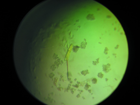
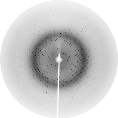
The rat protein tyrosine phosphatase eta (rPTPeta) is a cysteine-dependent phosphatase which hydrolyzes phosphoester bonds in proteins and other molecules. rPTPeta and its human homologue DEP-1 are involved in neoplastic transformations. Thus, expression of the protein is reduced in all oncogene-transformed thyroid cell lines and is absent in highly malignant thyroid cells. Moreover, consistent with the suggested tumour suppression role of PTPeta, inhibition of the tumorigenic process occurs after its exogenous reconstitution, suggesting that PTPeta might be important for gene therapy of cancers. In this study, the catalytic domain of rPTPeta was produced in Escherichia coli in soluble form and purified to homogeneity. Crystals were obtained by the hanging-drop vapour-diffusion method. Diffraction data were collected to 1.87 A resolution. The crystal belongs to space group P2(1)2(1)2(1), with unit-cell parameters a = 46.46, b = 63.07, c = 111.64 A, and contains one molecule per asymmetric unit.
Figures
Similar articles
-
Expression, purification, and crystallization of the catalytic domain of protein tyrosine phosphatase SHP-1.J Struct Biol. 1997 Nov;120(2):201-3. doi: 10.1006/jsbi.1997.3927. J Struct Biol. 1997. PMID: 9417985
-
Crystallization and preliminary X-ray analysis of the low molecular weight phosphotyrosyl protein phosphatase from bovine heart.J Mol Biol. 1994 Apr 29;238(2):281-3. doi: 10.1006/jmbi.1994.1287. J Mol Biol. 1994. PMID: 8158654
-
Purification and crystallization of the catalytic domain of human protein tyrosine phosphatase 1B expressed in Escherichia coli.J Mol Biol. 1994 Jun 24;239(5):726-30. doi: 10.1006/jmbi.1994.1409. J Mol Biol. 1994. PMID: 8014992
-
Crystallization and preliminary X-ray studies of cold-active protein-tyrosine phosphatase of Shewanella sp.Acta Crystallogr D Biol Crystallogr. 2002 Sep;58(Pt 9):1465-6. doi: 10.1107/S0907444902010302. Epub 2002 Aug 23. Acta Crystallogr D Biol Crystallogr. 2002. PMID: 12198303
-
Purification, crystallization and preliminary X-ray diffraction analysis of yeast nucleosome-assembly factor Cia1p.Acta Crystallogr D Biol Crystallogr. 2002 Oct;58(Pt 10 Pt 2):1876-8. doi: 10.1107/s0907444902013860. Epub 2002 Sep 28. Acta Crystallogr D Biol Crystallogr. 2002. PMID: 12351844
References
-
- Alonso, A., Sasin, J., Bottini, N., Friedberg, I., Friedberg, I., Osterman, A., Godzik, A., Hunter, T., Dixon, J. & Mustelin, T. (2004). Cell, 117, 699–711. - PubMed
-
- Ardini, E., Agresti, R., Tagliabue, E., Greco, M., Aiello, P., Yang, L. T., Ménard, S. & Sap, J. (2000). Oncogene, 19, 4979–4987. - PubMed
-
- Barford, D., Das, A. K. & Egloff, M. P. (1998). Annu. Rev. Biophys. Biomol. Struct.27, 133–164. - PubMed
-
- Brünger, A. T., Adams, P. D., Clore, G. M., DeLano, W. L., Gros, P., Grosse-Kunstleve, R. W., Jiang, J.-S., Kuszewski, J., Nilges, M., Pannu, N. S., Read, R. J., Rice, L. M., Simonson, T. & Warren, G. L. (1998). Acta Cryst. D54, 905–921. - PubMed
-
- Chagnon, M. J., Uetani, N. & Tremblay, M. L. (2004). Biochem. Cell Biol.82, 664–675. - PubMed
Publication types
MeSH terms
Substances
Associated data
- Actions
LinkOut - more resources
Full Text Sources
