. 2006 Sep 20;25(18):4253-62.
doi: 10.1038/sj.emboj.7601305. Epub 2006 Aug 31.

3'-end formation signals modulate the association of genes with the nuclear periphery as well as mRNP dot formation

Affiliations

3'-end formation signals modulate the association of genes with the nuclear periphery as well as mRNP dot formation

Katharine C Abruzzi et al. EMBO J. .

Abstract

Multiple studies indicate that mRNA processing defects cause mRNAs to accumulate in discrete nuclear foci or dots, in mammalian cells as well as yeast. To investigate this phenomenon, we have studied a series of GAL reporter constructs integrated into the yeast genome adjacent to an array of TetR-GFP-bound TetO sites. mRNA within dots is predominantly post-transcriptional, and dots are adjacent to but distinct from their transcription site. These reporter genes also localize to the nuclear periphery upon gene induction, like their endogenous GAL counterparts. Surprisingly, this peripheral localization persists long after transcriptional shutoff, and there is a comparable persistence of the RNA in the dots. Moreover, dot disappearance and gene delocalization from the nuclear periphery occur with similar kinetics after transcriptional shutoff. Both kinetics depend in turn on reporter gene 3'-end formation signals. Our experiments indicate that gene association with the nuclear periphery does not require ongoing transcription and suggest that the mRNPs within dots may make a major contribution to the gene-nuclear periphery tether.


Figures

Figure 1
Figure 1
FISH of cells expressing GFP reporter constructs. (A) Diagram of four GFP reporter constructs used in this study. All constructs contain the GFP open reading frame under the control of either the constitutive TDH3 promoter or the inducible GAL1 promoter. The 3′-end of the constructs has either the TDH3 or GAL1 3′UTR or a self-cleaving hammerhead ribozyme (RZ). The position of the GFP FISH probes is shown. (B) FISH with a GFP-specific probe shows that GFP mRNA with a ribozyme generated 3′-end are retained as dots within the nucleus (TDH-GFP-RZ and GAL-GFP-RZ). In contrast, TDH3-promoted GFP RNAs that contain a TDH3 pA signal are diffusely localized throughout the cell (TDH-GFP-TDHpA). Surprisingly, GAL1-promoted GFP mRNAs that are polyadenylated via a GAL1 pA signal are retained dots within the nucleus (GAL-GFP-GALpA).
Figure 2
Figure 2
Dot formation is dependent on the nature of the 3′UTR. (A) Diagram of the two chimeric GFP reporter constructs. These constructs contain the GFP open reading frame and have either a TDH3 promoter and a GAL1 3′UTR (TDH-GFP-GALpA) or a GAL1 promoter and a TDH3 3′UTR (GAL-GFP-TDHpA). The position of the GFP FISH probes is shown. (B) FISH with a GFP-specific probe (red) and DAPI staining (blue) of the DNA was performed on TDH-GFP-GALpA and GAL-GFP-TDHpA cells grown either in glucose (GLU) or galactose (GAL). TDH-GFP-GALpA mRNAs are expressed and retained in dots in the nucleus when cells are grown in either glucose or galactose. GAL-GFP-TDHpA mRNAs are not expressed in glucose and in galactose they are diffusely localized throughout the cell.
Figure 3
Figure 3
GAL-GFP-GALpA and GAL-GFP-RZ dots are independent of the nuclear exosome component, Rrp6p. Cells expressing integrated GAL-GFP-GALpA or GAL-GFP-RZ in either a WT or Δrrp6 background were grown in galactose and message specific FISH was performed using oligos specific to GFP (see Figures 1 and 2). The ability of GAL-GFP-GALpA and GAL-GFP-RZ dots to form is not dependent on Rrp6p.
Figure 4
Figure 4
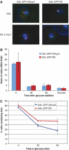
GFP dots are adjacent to but not overlapping the site of transcription and persist in the absence of transcription. (A) The GFP reporter genes were integrated adjacent to a TetO operator array to which TetR-GFP binds. When transcription is activated by steady-state growth in galactose, the GFP dots (FISH; shown in red) are adjacent to but not overlapping the site of transcription (TetR-GFP; shown in green). At 60 min after transcription is repressed by the addition of glucose, the dots are still visible and maintain a similar spatial relationship with the DNA locus. (B) Cells expressing GAL-GFP-GALpA or GAL-GFP-RZ were grown in galactose and at time zero, glucose was added to repress transcription. Cells were harvested for ChIP prior to glucose addition, 30 and 60 min after glucose addition and from cells maintained in glucose. RNA PolII occupancy on GAL-GFP-GALpA (blue) and GAL-GFP-RZ (red) was monitored at each timepoint. After 30 min, PolII levels are similar to those observed when transcription is repressed in glucose. (C) FISH using a GFP-specific probe was performed on the same cells used in (B). The number of cells containing GFP dots was counted (see Materials and methods). Both GAL-GFP-GALpA (blue) and GAL-GFP-RZ (red) expressing cells contain dots after transcription has been repressed by the addition of glucose. The GAL-GFP-RZ dots are more stable than the GAL-GFP-GALpA dots.
Figure 5
Figure 5
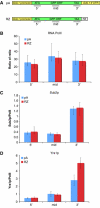
Co-transcriptional recruitment of mRNP proteins to GAL-GFP-GALpA and GAL-GFP-RZ. ChIPs were performed with RNA PolII and two different mRNP proteins (Sub2p and Yra1p) and the levels of each factor on GAL-GFP-GALpA and GAL-GFP-RZ was determined. (A) Diagram of the GFP reporters and the location of the PCR primers used in ChIP experiments. (B) RNA PolII recruitment is similar on GAL-GFP-GALpA and GAL-GFP-RZ. (C) The mRNP protein, Sub2p, is recruited similarly to both reporter genes. (D) Yra1p is enriched on the 3′-end of the GAL-GFP-RZ gene in comparison to the GAL-GFP-GALpA gene.
Figure 6
Figure 6
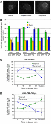
Intranuclear localization of the GAL-GFP-GALpA, GAL-GFP-RZ and GAL-GFP-TDHpA genes. The GAL reporter genes were integrated into the genome adjacent to a TetO array. TetR-GFP was co-expressed to visualize the gene and NUP49-CFP was expressed to mark the nuclear periphery. (A) The intranuclear localization of the gene (TetR-GFP) relative to the nuclear periphery was categorized as being internal (I), subperipheral (S; proximal to but not overlapping the nuclear membrane), or peripheral (P; adjacent to the nuclear membrane). Representative images of each category are shown. (B) The intranuclear localization of the GAL-GFP-GALpA, GAL-GFP-RZ and GAL-GFP-TDHpA genes was determined during transcriptionally active (GAL; galactose) or repressed (GLU; glucose) states. Both GAL-GFP-GALpA and GAL-GFP-RZ become more associated with the nuclear periphery when grown in galactose. In contrast, there was no substantial difference in the localization of the GAL-GFP-TDHpA gene in glucose compared to galactose. (C) The intranuclear localization of the GAL-GFP-RZ gene was examined 0, 15, 30 and 60 min after transcriptional shutoff by the addition of glucose as well as in steady-state glucose. The GAL-GFP-RZ gene is enriched in the nuclear periphery for up to 60 min after transcriptional shutoff suggesting that the GAL-GFP-RZ gene does not require active transcription to maintain its peripheral localization. (D) The intranuclear localization of the GAL-GFP-GALpA gene was examined 0, 15, 30 and 60 min after transcriptional shutoff by the addition of glucose as well as in steady-state glucose. The GAL-GFP-GALpA remains at the nuclear periphery for 30 min after transcriptional shutoff illustrating that the gene does not require active transcription to maintain its peripheral localization. However, the GAL-GFP-GALpA is released from the nuclear periphery more quickly than the GAL-GFP-RZ gene.
Figure 7
Figure 7
GAL1 mRNAs accumulate in dots that are independent of the nuclear exosome component, Rrp6p. WT or Δrrp6 cells were grown in galactose and message specific FISH was performed using oligos specific to GAL1. Dapi staining (blue), GAL1-specific FISH (red) and the overlapping signals are shown.
Figure 8
Figure 8
Model. (A) When GAL genes are actively transcribed, mRNPs accumulate in a dot that is adjacent to but not overlapping with the gene. Consistent with the observations of others, we show that GAL genes localize to the nuclear periphery and that a gene/nuclear periphery tether exists that is likely to include actively transcribing chromatin and its associated proteins. (B) When transcription of the GAL genes is repressed, dots persist and maintain the same spatial relationship with the gene. In addition, the gene remains associated with the nuclear periphery. These results suggest the existence of both a dot/gene and a gene/nuclear periphery tether that are independent of transcription. Both dot persistence and maintenance of nuclear periphery association are dependent on the 3′end (pA versus RZ). Although these differences could be affected by chromatin modifications that are maintained in the absence of transcription, we suggest that they are due to changes in the quantity or quality of the post-transcriptional mRNPs. It is also possible that post-transcriptional mRNPs also play a role in the gene/nuclear periphery tether during active transcription (see Discussion).

References

    1. Abruzzi KC, Lacadie S, Rosbash M (2004) Biochemical analysis of TREX complex recruitment to intronless and intron-containing yeast genes. EMBO J 23: 2620–2631 - PMC - PubMed
    1. Agard DA, Hiraoka Y, Shaw P, Sedat JW (1989) Fluorescence microscopy in three dimensions. Methods Cell Biol 30: 353–377 - PubMed
    1. Amberg DC, Fleischmann M, Stagljar I, Cole CN, Aebi M (1993) Nuclear PRP20 protein is required for mRNA export. EMBO J 12: 233–241 - PMC - PubMed
    1. Blobel G (1985) Gene gating: a hypothesis. Proc Natl Acad Sci USA 82: 8527–8529 - PMC - PubMed
    1. Brickner JH, Walter P (2004) Gene recruitment of the activated INO1 locus to the nuclear membrane. PLoS Biol 2: e342. - PMC - PubMed

Publication types

MeSH terms

LinkOut - more resources